Σαν δεν ντρέπονται, βγάζουν συμπέρασμα από ένα περιστατικό.

Για όνομα του Θεού, βιάζονται να μας εμβολιάσουν όλους στα γρήγορα και πάση θυσία! Το σχέδιο απομείωσης των λαών σε πλήρη δράση!

Πόσο επικίνδυνο είναι να εμβολιαστούμε αν νοσούμε από COVID-19; Τι έδειξε η πρώτη μελέτη περιστατικού

Πόσο επικίνδυνο είναι να εμβολιαστούμε αν νοσούμε από COVID-19; Τι έδειξε η πρώτη μελέτη περιστατικού

Ο ιός SARS-CoV-2, ο οποίος ενοχοποιείται για την πανδημία της COVID-19, συνεχίζει να εξαπλώνεται παγκοσμίως και έχει επηρεάσει ήδη πάνω από 160 εκατομμύρια άτομα, προκαλώντας σχεδόν 3 εκατομμύρια θανάτους.

Σχεδόν 1 έτος μετά το πρώτο περιστατικό της πανδημίας το Δεκέμβριο του 2019, οι περισσότερες χώρες έχουν ήδη ξεκινήσει τον εμβολιασμό του πληθυσμού τους.

Αναπόφευκτα, κάποια άτομα που θα κάνουν το εμβόλιο για την COVID-19, θα έχουν ενεργό λοίμωξη (συνήθως εν αγνοία τους).

Τι συμβαίνει σε αυτή την περίπτωση;

Είναι επικίνδυνο να κάνει το εμβόλιο κάποιος που νοσεί, ακόμα και με ασυμπτωματική λοίμωξη, από COVID-19;

Μία ομάδα επιστημόνων εξέτασε το περιστατικό ενός 31χρονου γιατρού από τις ΗΠΑ, θέλοντας να δώσει απαντήσεις στα παραπάνω ερωτήματα.

Το περιστατικό δημοσιεύτηκε στο επιστημονικό περιοδικό Infection και δίνει ορισμένες σημαντικές πληροφορίες για τη διαχείριση αντιστοίχων καταστάσεων.

Εμβολιασμός για την COVID-19

Από το Δεκέμβριο του 2020, αρκετές χώρες έχουν ξεκινήσει τον εμβολιασμό του πληθυσμού τους, δίνοντας προτεραιότητα στους ηλικιωμένους και τους εργαζομένους στον τομέα της υγείας.

Σταδιακά, ξεκίνησε και ο εμβολιασμός άλλων πληθυσμιακών ομάδων, με σκοπό να επιτευχθούν όσο το δυνατόν υψηλότερα ποσοστά ανοσίας“, σημειώνει ο κ. Αντώνιος Δημητρακόπουλος – Διευθυντής Γ’ Παθολογικής Κλινικής Ερρίκος Ντυνάν Hospital.

Η τεχνολογία του mRNA

Η τεχνολογία του mRNA αποτελεί μία από τις νεότερες προσεγγίσεις στην ανάπτυξη εμβολίων και έχει χρησιμοποιηθεί σήμερα στα εμβόλια της Pfizer και της Moderna.

Η οδηγία που περιέχουν τα εμβόλια mRNA

Τα εμβόλια αυτά περιέχουν την οδηγία για την παραγωγή μίας πρωτεΐνης του ιού SARS-CoV-2.

Η οδηγία αυτή μεταφράζεται από τα κύτταρα του ανθρώπου, τα οποία ακολούθως παρουσιάζουν την πρωτεΐνη στην εξωτερική τους μεμβράνη, γεγονός που εκκινεί την ανοσιακή απόκριση γι’ αυτή την πρωτεΐνη“, εξηγεί ο κ. Δημητρακόπουλος, προσθέτοντας:

Στη φυσική λοίμωξη από τον ιό SARS-CoV-2, δεν γνωρίζουμε σήμερα όλα τα χαρακτηριστικά της ανοσιακής απόκρισης, ωστόσο φαίνεται ότι η μέση διάρκεια που απαιτείται για την ανίχνευση IgG όσο και IgM αντισωμάτων είναι περίπου 2 εβδομάδες από την εμφάνιση των συμπτωμάτων“.

Προφανώς, η διάρκεια αυτή μπορεί να επηρεαστεί από διάφορους παράγοντες, όπως για παράδειγμα τη σοβαρότητα των συμπτωμάτων.

Το περιστατικό

Στη μελέτη τους, οι επιστήμονες εξέτασαν το περιστατικό ενός άνδρα γιατρού ο οποίος παρουσίασε συμπτώματα COVID-19,  λίγο μετά την 1η δόση του εμβολίου.

Ο 31χρονος άνδρας εργαζόταν σε μία κλινική με ασθενείς COVID-19 και εμφάνισε συμπτώματα που ομοιάζουν γρίπη λίγες ώρες μετά την 1η δόση του εμβολίου της Pfizer/BioNTech.

Ο ασθενής ανέφερε συμπτώματα όπως:

-κεφαλαλγία,

-βήχα,

-ρίγος,

-πυρετό και,

-γενικευμένο άλγος στα άκρα.

Όταν διαπίστωσε ότι τα συμπτώματά του δεν υποχώρησαν μετά τις πρώτες 24 ώρες, έκανε εξετάσεις για τον SARS-CoV-2 οι οποίες βγήκαν θετικές.

Καθώς τα αναπνευστικά του συμπτώματα δεν ήταν σοβαρά, του ζητήθηκε να παραμείνει σε καραντίνα στο σπίτι.

“Καθώς ο ασθενής είχε κάνει την 1η δόση του εμβολίου mRNA και είχε θετικές εξετάσεις για την COVID-19 σε μικρό χρονικό διάστημα, το περιστατικό του είχε ιδιαίτερο ενδιαφέρον“, αναφέρει ο κ. Δημητρακόπουλος, προσθέτοντας:

Θέλοντας να εξετάσουν αν τα συμπτώματά του αποτελούν ανεπιθύμητες ενέργειες του εμβολίου ή αποδίδονται στον ιό SARS-CoV-2 οι επιστήμονες της παρούσας μελέτης ασχολήθηκαν αρκετά με το συγκεκριμένο περιστατικό“.

Θέλησαν επίσης να διαπιστώσουν αν τα mRNA εμβόλια μπορεί να επηρεάσουν την ακρίβεια της εξέτασης PCR, καθώς και αν ο εμβολιασμός κατά τη διάρκεια ενεργού λοίμωξης είναι επικίνδυνος για τον ασθενή.

Για να απαντήσουν στα παραπάνω ερωτήματα, οι επιστήμονες εξέτασαν την απόκριση αντισωμάτων του ασθενούς.

Στο ρινοφαρυγγικό δείγμα που έλαβαν μετά τη διάγνωση της νόσου, τα επίπεδα του ιού είχαν περιοριστεί ήδη σε 1.7 εκατομμύρια αντίγραφα/ml, από τα 32 εκατομμύρια/ml της πρώτης εξέτασης.

Όταν η επιστημονική ομάδα επανέλαβε την εξέτασε μετά από 10 ημέρες, το ιικό φορτίο είχε μειωθεί ακόμα περισσότερο και είχε φτάσει στα 0.013 εκατομμύρια αντίγραφα/ml.

Ο ασθενής είχε αντισώματα IgG τόσο για την πρωτεΐνη του νουκλεοκαψιδίου, όσο και την πρωτεΐνη ακίδα.

Συμπεράσματα

Οι επιστήμονες κατέληξαν ότι οι εργαζόμενοι σε περιβάλλον με υψηλό κίνδυνο έκθεσης στον ιό έχουν αυξημένη πιθανότητα να έχουν ενεργό λοίμωξη COVID-19 όταν θα έρθει η ώρα να εμβολιαστούν.

Επιπλέον, καθώς το εμβόλιο χορηγείται στο δελτοειδή μυ, η πιθανότητα θετικής εξέτασης PCR από ρινοφαρυγγικό δείγμα είναι πολύ χαμηλή“, τονίζει ο κ. Δημητρακόπουλος.

Το σημαντικό σημείο

Το σημαντικό σημείο στο οποίο στάθηκαν οι επιστήμονες της έρευνας ήταν ότι το εμβόλιο είχε καλή ανοχή, παρά το γεγονός ότι ο γιατρός είχε ήδη ενεργό λοίμωξη από SARS-CoV-2.

Όταν οι επιστήμονες εξέτασαν τα επίπεδα της ιντερλευκίνης-6, της CRP, της προκαλσιτονίνης, των λευκοκυττάρων και των δεικτών του ήπατος και των νεφρών, δεν παρατήρησαν κανένα παθολογικό εύρημα 7 ημέρες μετά την εμφάνιση των συμπτωμάτων“, εξηγεί ο κ. Δημητρακόπουλος.

Με βάση τα παραπάνω, δεν υπάρχει ανάγκη να γίνονται εξετάσεις για τον ιό πριν τη χορήγηση του εμβολίου, καθώς ο εμβολιασμός κατά τη διάρκεια της λοίμωξης δεν φαίνεται να συνδέεται με χειρότερη πρόγνωση.

Καταλήγοντας, οι επιστήμονες υποστήριξαν ότι οποιοσδήποτε κάνει εμβόλιο και έχει ανεπιθύμητες ενέργειες για περισσότερο από 1 ημέρα, θα πρέπει ίσως να κάνει rapid test.

ΠΗΓΗ:https://www.ygeiamasnews.gr/astheneies/epidimies/54868/poso-epikindyno-einai-na-emvoliastoume-an-nosoume-apo-covid-19-ti-edeikse-i-proti-meleti-peristatikou/

Θα εμβολιάσουν, δηλαδή θα αφαιρέσουν κάμποσες ζωές από την ΕΜΑΚ οι εκτελεστές-φερέφωνα του σχεδίου της χαζαρικής παγκοσμιοποίησης

Υποχρεωτικός ο εμβολιασμός για όσους υπηρετούν στην ΕΜΑΚ

Υποχρεωτικός ο εμβολιασμός για όσους υπηρετούν στην ΕΜΑΚ

Υποχρεωτικό εμβολιασμό του συνόλου των πυροσβεστών που υπηρετούν στην ΕΜΑΚ προβλέπει διαταγή που εξέδωσε την Τρίτη το Αρχηγείο της Πυροσβεστικής Υπηρεσίας.

Η διαταγή εκδόθηκε με πρωτοβουλία και απόφαση του ίδιου του Αρχηγού του Σώματος Στέφανου Κολοκούρη, που υπήρξε επικεφαλής της συγκεκριμένης υπηρεσίας.

Ανώτατος αξιωματικός του Σώματος που ρωτήθηκε σχετικά διευκρίνισε ότι η ΕΜΑΚ ως ειδική υπηρεσία συμμετέχει στον ευρωπαϊκό μηχανισμό πολιτικής προστασίας και ως εκ τούτου οφείλει ανά πάσα στιγμή να είναι έτοιμη να συμμετάσχει -κατόπιν αιτήματος της Ε.Ε. σε επιχειρήσεις διάσωσης σε όλο των κόσμο.

Ως εκ τούτου το προσωπικό της οφείλει να είναι εμβολιασμένο τόσο για κορωνοϊό όσο και για άλλες μεταδοτικές ασθένειες προκειμένου να είναι σε θέση να ανταποκριθεί στα καθήκοντά του.

Όσοι δεν θελήσουν να εμβολιαστούν θα μετακινηθούν σε άλλη υπηρεσία της Π.Υ.

Αυτή είναι η διαταγή που εξέδωσε σήμερα το Αρχηγείο της Πυροσβεστικής Υπηρεσίας και προβλέπει υποχρεωτικό εμβολιασμό του συνόλου των πυροσβεστών που υπηρετούν στην ΕΜΑΚ.

Πηγή: ΕΡΤ

COVID-19 is not “just another flu” but a quite severe disease in comparison of influenza A and B

COVID-19 is not “just another flu”: a real-life comparison of severe COVID-19 and influenza in hospitalized patients in Vienna, Austria

Team members

Abstract

Background

COVID-19 is regularly compared to influenza. Mortality and case-fatality rates vary widely depending on incidence of COVID-19 and the testing policy in affected countries. To date, data comparing hospitalized patients with COVID-19 and influenza is scarce.

Methods

Data from patients with COVID-19 were compared to patients infected with influenza A (InfA) and B (InfB) virus during the 2017/18 and 2018/19 seasons. All patients were ≥ 18 years old, had PCR-confirmed infection and needed hospital treatment. Demographic data, medical history, length-of-stay (LOS), complications including in-hospital mortality were analyzed.

Results

In total, 142 patients with COVID-19 were compared to 266 patients with InfA and 300 with InfB. Differences in median age (COVID-19 70.5 years vs InfA 70 years and InfB 77 years, p < 0.001) and laboratory results were observed. COVID-19 patients had fewer comorbidities, but complications (respiratory insufficiency, pneumonia, acute kidney injury, acute heart failure and death) occurred more frequently.

Median length-of-stay (LOS) was longer in COVID-19 patients (12 days vs InfA 7 days vs. InfB 7 days, p < 0.001). There was a fourfold higher in-hospital mortality in COVID-19 patients (23.2%) when compared with InfA (5.6%) or InfB (4.7%; p < 0.001).

Conclusion

In hospitalized patients, COVID-19 is associated with longer LOS, a higher number of complications and higher in-hospital mortality compared to influenza, even in a population with fewer co-morbidities. This data, a high reproduction number and limited treatment options, alongside excess mortality during the SARS-CoV-2 pandemic, support the containment strategies implemented by most authorities.

Introduction

As of July 26th 2020, the ongoing SARS-CoV-2 pandemic has infected over 68 million people and has caused over 1,560,000 deaths worldwide [1], challenging affected countries and healthcare systems. The pandemic has impacted the lives of billions on multiple levels and has had a major impact on the world economy [2].

Influenza viruses are highly contagious and during seasonal epidemics excess mortality is observed. While therapeutic options and vaccinations exist for influenza vaccination rollout for COVID-19 is still limited and delayed by supply problems, and whilst there is growing evidence that certain drugs may be effective, these are still deemed to have limited therapeutic effect [3,4,5,6,7,8,9,10]. In comparison to COVID-19 there are no strict regulations and containment strategies during seasonal influenza virus epidemics.

While mortality and case-fatality-rates of COVID-19 seem to be higher than for influenza A (InfA) and influenza B (InfB), reliable information is difficult to obtain due to asymptomatic and oligosymptomatic presentations in both cases. Asymptomatic cases are described in 18–75% for SARS-CoV-2 [11, 12]) and 4–85% for influenza [13]. Case-fatality rate estimates range from 0.25 to 5.7% for COVID-19 [14, 15] and from 0.1 to 1% for influenza [16].

Here we compare the demographic data, medical history, length-of-stay (LOS), complications including ICU admission and in-hospital mortality between hospitalized PCR confirmed patients with COVID-19, InfA and InfB. Treatment and isolation were performed at the same department by the same specialists for infectious diseases, ensuring equal quality of medical care.

Methods

Study design and data gathering

This study was conducted at the Department for Infectious Diseases and Tropical Medicine at the Kaiser-Franz-Josef Hospital in Vienna, Austria. We compared demographics, medical history, laboratory results, LOS and complications including ICU admission and in-hospital mortality of patients with polymerase chain reaction (PCR)-proven COVID-19, InfA and InfB virus infections.

Data from COVID-19 patients were collected from March 1st to April 25th 2020. PCR testing for SARS-CoV-2 took place at our hospital’s laboratory institute or at other certified laboratories in Vienna. Data from all subsequent influenza patients were collected retrospectively in 2017/18 and prospectively in 2018/19. Influenza diagnosis was made at the emergency department before admission using the Alere™ i Influenza A & B assay (Alere, Waltham, MA, USA) in 2017/18 and the Cobas® Liat® point-of-care test (POCT) from Roche (Roche Molecular Systems, Pleasanton, CA, USA) in 2018/19.

All hospitalized patients ≥ 18 years with molecular proven COVID-19 or influenza were eligible for the study. Patients’ medical history, laboratory parameters and complications were collected via a standardized form during hospital admission. Incomplete data were updated retrospectively from patients’ electronic health records whenever possible.

The study was approved by the local ethics committee.

Definition of variables

The first day of any COVID-19 or influenza-associated symptom was considered to be disease onset. Fever was defined as a body temperature ≥ 38 °C. Dehydration was defined as the need for intravenous fluids based on clinical appearance. Respiratory insufficiency was defined as SpO2 ≤ 93% at room air or the need for supplementary oxygen based on clinical judgment by the treating physician. Pneumonia was defined as typical consolidation and/or opacity on a radiological image. Myositis was defined as a creatine-kinase (CK) level above 1000 U/L. Heart failure was defined by new onset or worsening of peripheral edema and/or congestion on X-ray in patients with history of chronic heart failure and without any other cause. Acute kidney injury was defined as either an increase of creatinine level by 0.3 mg/dl from the baseline kidney function within 48 h or an increase of ≥ 1.5 times the baseline (presumed to have occurred within the previous 7 days due to the current episode of illness). When no previous creatinine level was available as baseline, the acute kidney injury was assessed retrospectively. We did not differentiate between complications which were present on admission or developed during admission.

Statistical analysis

Data were double-checked, entered in a MS Excel sheet (Microsoft, Redmond, WA, USA) and anonymized before statistical analysis. The statistical analyses were performed with SAS V9.4 and R Version4.0.2. Categorical variables were described by counts and percentages. For metric non-normally distributed variables the median (Md) and interquartile range (IQR) were used. Significance tests for categorical variables were made via cross tables and Chi-squared test or Fisher’s exact test, where applicable. Kruskal–Wallis tests were performed to compare the three groups (InfA, InfB and COVID-19) for metric non-normally distributed variables. A two-sided alpha < 0.05 was considered statistically significant.

To check for difference in binary outcomes (e.g. acute heart failure, in-hospital mortality) logistic regression analyses were performed with the factor group (InfA, InfB vs COVID-19). Odds ratios and two-sided 95% confidence intervals reported.

For in-hospital mortality and discharged alive from hospital analysis, competing risk analysis was performed and the cumulative incidence curves were compared using Gray’s test (Gray 1988).

Results

Patients demographics and medical history

The total population consisted of 708 patients, 142 (20.1%) had COVID-19, 266 (37.6%) InfA and 300 (42.3%) InfB. 356 (50.3%) were male, with a higher proportion of male patients in the COVID-19 group. Overall median age was 73.5 years (61–82) and varied between groups (p < 0.001); overall InfB patients were oldest.

Differences in medical history were demonstrated between the groups, with lower rates of chronic kidney disease (p < 0.001) and chronic obstructive disease (p = 0.002) in the COVID-19 group. Median time from symptom onset to hospitalization was 7 days (IQR 3–10) in the COVID-19 group and differed significantly from InfA (2 days; IQR 1–4) and InfB (2 days; IQR 0.8–4) positive patients (p < 0.001). Antiviral treatment differed significantly between groups (p < 0.001), while antibiotic prescription rates did not yield a statistical significance (p = 0.11). For details see Table 1.


Table 1 Patients demographics and medical history

From: COVID-19 is not “just another flu”: a real-life comparison of severe COVID-19 and influenza in hospitalized patients in Vienna, Austria

Total
(n = 708)
SARS-CoV-2
(n = 142)
Influenza A
(n = 266)
Influenza B
(n = 300)
p value
Age (years)a 73.5 (61–82) 70.5 (53–80) 70 (58–80) 77 (67–85)  < 0.001
Sex (male) 356/708 (50.3%) 84 (59.2%) 131 (49.3%) 141 (47%) 0.053
BMI (kg/m2) 25.6 (22.7–30.1) [n = 488]b 25.9 (24.2) [n = 75] 25.3 (22.2–29.7) [n = 218] 26 (23–30.5) [n = 195] 0.246
Time from symptom onset to hospitalization 3 days (1–5) n = 616 7 days (3–10) n = 126 2 days (1–4) n = 244 2 days (0.8–4) n = 246  < 0.001
Antiviral treatment 433 (61.1%) 67 (47.2%)c 188 (70.7%)d 178 (59.3%)d  < 0.001
Antibiotic treatment 282 (39.8%) 46 (32.4%) 114 (42.9%) 122 (40.7%) 0.112
Medical history
 Chronic kidney disease 212 (29.9%) 26 (18 .3%) 75 (28.2%) 111 (37%)  < 0.001
 Obstructive pulmonary disease 194 (27.4%) 23 (16.2%) 92 (34.6%) 79 (26.3%)  < 0.001
 Diabetes 176 (24.9) 27 (19%) 69 (25.9%) 80 (26.7%) 0.193
 Atrial fibrillation 133 (18.8%) 30 (21.1%) 46 (17.3%) 57 (19%) 0.635
 Coronary heart disease 48/312 (15.4%) 20/142(14.1%) 28/170 (16.5%) NA 0.56
 Any malignancy 94 (13.3%) 12 (8.5%) 31 (11.7%) 51 (17%) 0.029
 Dementia 82 (11.6%) 12 (8.5%) 30 (11.3%) 40 (13.3%) 0.142
 Congestive heart failure 75 (10.6%) 17 (12%) 28 (10.5%) 30 (10%) 0.82
 Peripheral artery disease 45/707 (6.4%) 7/141 (5%) 14 (5.3%) 24 (8%) 0.309
 Rheumatic disease 11/312 (3.5%) 6 (4.2%) 5/170 (2.9%) NA 0.54
  1. Significant differences are marked in bold
  2. NA not available (data were not available in this subgroup)
  3. aMedian and interquartile range are shown
  4. bIf data were not available for all patients, the number of valid observations per variable and group is additionally reported with n = for numeric data as ratio x/n for binary data in the respective cell
  5. c44 patients received lopinavir/ritonavir, 18 hydroxychloroquine, one both drugs, one lopinavir/ritonavir plus remdesivir, three camostat; oseltamivir
  6. dOseltamivir was used as antiviral treatment

    Laboratory parameters on admission

    All laboratory parameters except creatine-kinase (p = 0.115) and bilirubin (p = 0.062) differed significantly between groups. COVID-19 patients had the lowest leukocyte count (p < 0.001), highest C-reactive-protein (p < 0.001) and lactate-dehydrogenase level (p < 0.001). For further details see Table 2.

    Table 2 Laboratory parameters on admission

    From: COVID-19 is not “just another flu”: a real-life comparison of severe COVID-19 and influenza in hospitalized patients in Vienna, Austria

    Name (unit) [n] Total
    (n = 708)
    SARS-CoV-2
    (n = 142)
    Influenza A
    (n = 266)
    Influenza B
    (n = 300)
    p value
    Leukocytes (G/L) 6.8 (5–9.2) n = 704 5.9 (4.5–7.4) n = 139 7.7 (5.8–10.1) n = 265 6.8 (5–9.2)  < 0.001
    Platelets (G/L) 194 (149–249) n = 704 200 (155–255) n = 139 199 (158–256) n = 265 185 (143–238) 0.030
    C-reactive protein (mg/L) 34.3 (14.9–72.8) n = 702 60 (27.9–95.7) n = 137 40 (21–77) n = 265 23 (10–54)  < 0.001
    Creatinine (mg/dl) 1.00 (0.8–1.3) n = 700 1 (0.8–1.3) n = 138 0.93 (0.73–1.19) n = 263 1.04 (0.85–1.39) n = 299  < 0.001
    CK (U/L) 117 (65–223) n = 630 104 (52–202) n = 124 119 (62–246) n = 242 119 (73–214) n = 264 0.115
    ALT (U/L) 28 (18–40) n = 339 31 (19–43) n = 125 25 (17–34) n = 174 38 (24–57) n = 40  < 0.001
    Bilirubin (mg/dl) 0.5 (0.3–0.7) n = 643 0.5 (0.35–0.69) n = 131 0.45 (0.28–0.63) n = 242 0.45 (0.31–0.66) n = 270 0.062
    LDH (U/L) 245 (205–312) n = 453 303 (234–382) n = 121 229 (194–282) n = 165 233 (203–289) n = 165  < 0.001
    Troponin T-hs (ng/L) 18 (8.3–43.2) n = 174 13 (7–28) n = 94 22 (13–47) n = 45 26 (9–69) n = 35 0.005
    1. Not every information was available for each patient on admission, in such cases the number of valid observations per variable and group is reported with n = in the respective cell
    2. Significant differences are marked in bold
    3. IQR interquartile range, CK creatinine-kinase, ALAT alanine-amino-transferase, GGT gamma-glutamyl-transferase, AP alkalic phosphatase, LDH lactate-dehydrogenase, eGFR estimated glomerular-filtration-rate ap-values derive from Kruskal–Wallis test

      Complications and outcome

      COVID-19 infected patients had a significantly higher rate of respiratory insufficiency (COVID-19 66.9% vs 36.8% InfA vs 18.3% InfB, p < 0.001), pneumonia (COVID-19 76.1% vs 26.3% InfA vs 24% InfB, p < 0.001), acute kidney injury (COVID-19 27.5% vs 10.9% InfA vs 13.8% InfB, p < 0.001) and acute heart failure (COVID-19 10.6% vs 4.1% InfA vs 3.7% InfB, p = 0.006). The myositis rate was higher in influenza patients (InfA: 7.7%, InfB 8.1%) compared to COVID-19 (3.5%), but this was not statistically significant (p = 0.19) and the rate of acute coronary syndrome did not differ between groups. Odds ratios and 95% confidence intervals are presented in Fig. 1.

      Fig. 1

      From: COVID-19 is not “just another flu”: a real-life comparison of severe COVID-19 and influenza in hospitalized patients in Vienna, Austria

      Fig. 1

      Complications and outcome. Data are expressed as inverse OR. OR > 1, therefore, means the risk for the complication is higher in the SARS-CoV-2 group (e.g.: an OR of 6.25 in the InfB line for in-hospital mortality means the risk for in-hospital mortality is 6.25 times higher for COVID-19 patients. ICU intensive care unit

    In-hospital mortality differed significantly between groups with 23.2% for COVID-19, 5.6% for InfA and 4.7% for InfB (p < 0.001). COVID-19 had a higher in-hospital mortality compared to InfA (OR 5; 95% CI 2.63–10) and InfB (OR 6.25; 95% CI 3.23–12.5), see Fig. 1. The cumulative incidence curves for in-hospital mortality and discharge from hospital also yielded statistically significant differences (see Fig. 2). The median time to patients being discharged from hospital (length of stay, LOS) is almost double for COVID-19 patients when compared to those with InfA and InfB (12 days vs 7 days, respectively). The groups did not reach statistically significant differences with respect to ICU admission rates (COVID-19 8.5% vs InfA 9% vs InfB 4.3%, p = 0.065). For further details see Fig. 1.

  7. Fig. 2

    From: COVID-19 is not “just another flu”: a real-life comparison of severe COVID-19 and influenza in hospitalized patients in Vienna, Austria

    Fig. 2

    Cumulative incidence curves for in-hospital mortality and discharged-alive from hospital

    Mortality was strongly associated with older age in all groups. No patient under 60 died due to COVID-19 or influenza. Difference in in-hospital mortality increased with older age and was 2–13 times higher for COVID-19 compared to InfA or InfB when analyzing the age groups 61–70 years, 71–80 years, 81–90 years and > 90 years. For details see Table 3 and Fig. 3.

    Table 3 Age group-specific in-hospital mortality rate and ICU admission rate

    From: COVID-19 is not “just another flu”: a real-life comparison of severe COVID-19 and influenza in hospitalized patients in Vienna, Austria

     < 60 years 61–70 years 71–80 years 81–90 years  > 90 years Total
    In-hospital mortality
    SARS-CoV-2 0/47 (0%) 2/23 (8.7%) 8/38 (21.1%) 18/28 (64.3%) 5/6 (83.3%) 33/142 (23.2%)
    Influenza A 0/77 (0%) 3/61 (4.9%) 2/62 (3.2%) 7/55 (12.7%) 3/11 (27.3%) 15/266 (5.6%)
    Influenza B 0/48 (0%) 2/50 (4%) 3/94 (3.2%) 5/84 (6%) 4/24 (16.7%) 14/300 (4.7%)
    ICU admission rate
    SARS-CoV-2 5/42 (10.6%) 3/23 (13%) 4/38 (10.5%) 0/28 (0%) 0/6 (0%) 12/142 (8.5%)
    Influenza A 9/77 (11.7%) 8/61 (13.1%) 4/62 (6.5%) 3/55 (5.5%) 0/11 (0%) 24/266 (9%)
    Influenza B 5/48 (10%) 1/50 (2%) 4/94 (4.3%) 3/84 (3.6%) 0/24 (0%) 14/300 (4.3%)
    Fig. 3
    figure3

    Age group-specific in-hospital mortality rate

    Age group specific ICU admission rates are shown in Table 3. About 10% of patients under 60 years were admitted to the ICU. The rate decreased with higher age.

    Antimicrobial therapy

    The use of antiviral treatment did differ significantly between COVID-19, influenza A and influenza B (COVID-19 47.2%, InfA 70.7%, InfB 59.3%, p ≤ 0.001). While antiviral therapy was consistent within the influenza groups (only oseltamivir was used n = 366), antiviral treatment in COVID-19 patients varied (lopinavir/ritonavir n = 44/65.7%, hydroxychloroquine n = 18/26.7%, camostat n = 3/4.5%, lopinavir/ritonavir plus hydroxychloroquine n = 1/1.5%, lopinavir/ritonavir plus remdesivir n = 1/1.5%).

    There was no statistically significant difference in the use of antibiotic treatment (COVID-19 32.4%, InA 42.9%, InfB 40.7%, p = 0.112).

    Discussion

    In this study, we compared data from patients with severe COVID-19 and InfA and InfB, showing much higher in-hospital mortality for COVID-19. When comparing the patients’ demographics and medical history it was noted that InfB patients were significantly older. There was no statistically significant age difference between the COVID-19 and InfA group. The COVID-19 group had fewer comorbidities. Differences between InfA and InfB are already described in detail elsewhere and will not be discussed here [17]. Time from symptom onset to hospitalization was significantly longer in COVID-19 than in Influenza. This is in accordance with observations demonstrating that some SARS-CoV-2 infected patients deteriorate in the second week following symptom onset [18,19,20].

    Despite fewer comorbidities in the COVID-19 group, in-hospital mortality was much higher than in InfA or InfB (COVID-19 23.2%, InfA 5.6% and InfB 4.7%). During the time this study was conducted, there was no shortage of ICU beds or healthcare system overburden in Austria. The department for infectious diseases was additionally equipped with further AIRVO™ 2 humidification systems at the start of the pandemic to offer high-flow nasal oxygen therapy for patients on the normal ward which could have prevented ICU admissions. This was not the case during previous influenza virus epidemics. Other than that quality of medical care and therapeutic options was identical and, therefore, this should not have had a negative effect on COVID-19 in-hospital mortality. In accordance with latest scientific findings at the time COVID-19 patients were treated with lopinavir/ritonavir or hydroxychloroquine as off-label therapy if they met certain severity and safety criteria, while a higher proportion of influenza patients received specific antiviral treatment (Oseltamivir). Low-molecular-weight-heparin was given to all patients without contraindication for thromboembolic prophylaxis in all study groups. At the time of the study data regarding therapy was scarce and dexamethasone, tocilizumab, remdesivir, APN01 and convalescent plasma were only used at the ICU as part of clinical trials. In a further analysis different age groups were compared. In-hospital mortality is higher in the COVID-19 group in all age groups above the age of 60 years, and while older age is a well-described risk factor for all of the studied viral infections [19, 21] differences in mortality increase dramatically with older age (see Table 3 and Fig. 3). While InfA and InfB in-hospital mortality increased moderately when comparing age groups 61–70 years and 81–90 years this was much more pronounced in COVID-19. Older age is already described as a major risk factor for COVID-19 but seems to be of greater importance than in other viral infections [19, 20, 22] Possible explanations include immunosenescence and decreased immune responses for this novel coronavirus as opposed to immunological memory to previous antigen exposure in the case of influenza (original antigenic sin) [23, 24]. We did not collect data on influenza vaccinations which may have prevented additional deaths in older patients with influenza.

    While in-hospital mortality for influenza is widely known and accepted, recent publications demonstrate a significant increase when looking at 90-day mortality [25]. There is no information yet regarding long-term effects of COVID-19 or whether 30-day or 90-day mortality might be even higher than previously thought.

    Excess mortality is an important endpoint to measure the impact of diseases or other environmental phenomena on a large scale. While seasonal excess mortality associated with influenza seasons is well-acknowledged [7] data for the current pandemic only became available recently. Concerns arose that under-treatment of other medical issues and delay of urgent medical procedures might lead to increased non-COVID-19-related mortality. Recent data demonstrates a significant rise in excess mortality for Europe after the seasonal influenza epidemic during the time of the SARS-CoV-2 pandemic. This excess mortality is driven solely by countries with high numbers of COVID-19 cases. In Austria, where a strict and early containment strategy was practiced no excess mortality was observed [26].

    ICU admission rate did not differ in patients under 60 years of age. In the age group 61–70 years ICU admission rate was higher in the COVID-19 and InfA group. In addition, in the age group 71–80 years ICU admission was more common for COVID-19. In the COVID-19 group no patient older than 80 years was transferred to the ICU. Patients 80 years or older could either be managed with high-flow nasal oxygen therapy on the normal ward or ICU treatment was deemed to be non-beneficial for the patient due to comorbidities. Some patients also rejected ICU treatment on a personal decision. Transfer to ICU was not limited by availability during the pandemic in Austria. At our hospital the availability of high-flow oxygen on the normal ward did certainly reduce the number of ICU admissions. Unfortunately, as a downside of the fast and unbureaucratic initiation of high-flow oxygen on the normal ward as a response to the pandemic the documentation of high-flow oxygen usage was not standardized at the beginning and reliable interpretation to which extend this effected ICU admissions can therefore not be made.

    Of interest, men seemed to be disproportionately affected with COVID-19 as compared to patients hospitalized with Influenza. Higher rates of COVID-19-associated morbidity and mortality in men versus women were reported previously and could potentially be explained by higher prevalence of smoking and alcohol consumption as well as other comorbidities in men compared to women. However, no such difference was observed in Influenza. Among other factors, different expression patterns between men and women of the viral receptors for SARS-CoV2 but not for Influenza may explain these differences [27, 28].

    Alongside the higher mortality rate in COVID-19 other complications also appear to be more common. There was a significantly higher incidence of respiratory insufficiency, pneumonia, acute kidney injury and acute heart failure, as well as a significantly longer LOS which increases cost and burden on healthcare systems. The median time to patients being discharged from hospital is almost double for COVID-19 patients when compared to those with InfA and InfB (12 days vs 7 days respectively). The longer LOS might be partially explained due to hygienic reasons. At the beginning of the pandemic isolation of potentially infectious patients was particularly strict and two negative SARS-CoV-2 nasopharyngeal swaps have been necessary to end quarantine. Although people could theoretically have been discharged into self-quarantine at home or send to special “isolation-centers” this regularly delayed discharge, especially in people who needed personal assistance with activities of daily life. LOS for COVID-19 patients should be re-evaluated in further studies as rules for quarantine have changed considerably with increased knowledge about infectiousness and transmission of SARS-CoV-2 and adaption of care facilities to the challenges of the SARS-CoV-2 pandemic. Acute kidney injury was mostly mild and dialysis was not necessary in any patients on the normal ward, both in influenza patients as well as in COVID-19 patients. Organ replacement therapy as well as vasopressor use at the ICU were not analyzed in this study.

    Laboratory results differed significantly, but differentiation based on laboratory results is not possible. Most notable in our opinion is the higher C-reactive protein (CRP) level accompanied by a lower leucocyte count. This supports our clinical impression of high CRP levels in COVID-19 with viral origin while bacterial superinfections seem rare [20]. In InfA and InfB high CRP levels are often associated with bacterial superinfections [29,30,31]. Unfortunately, we were unable to compare lymphocyte counts and other laboratory results associated with bad prognosis in COVID-19 due to missing values in our influenza A and B data set.

    The strength of our study is that all patients had PCR-proven infections and were treated at the same department for infectious diseases by the same specialists, therefore, complications and in-hospital mortality could not have been influenced by different quality of healthcare or differences in expertise. The associated ICU was also managed by the same department. Successful containment strategies in Austria prevented ICU shortage during the time the data were collected which could have had an effect on mortality. To date, data comparing COVID-19, InfA and InfB in an elderly hospitalized population is scarce.

    The retrospective collection of some of the data is a limitation of our study. The age group-specific mortality estimates may be biased by the small sample size within some subgroups. Data were collected at a single institution and findings might not be applicable to other settings. Furthermore, this study only represents COVID-19 patients during the “first wave” in Austria. During the course of the pandemic management of COVID-19 patients, therapeutic options, vaccination rollout, public restrictions and behavior as well as mutations of the virus changed continuously and will continue doing so. The impact of these factors on public health systems, illness presentation and course as well as on demographic changes of COVID-19 patients is of great interest and needs continuous analysis and interpretation.

    In conclusion we could demonstrate the severity and high in-hospital mortality of COVID-19 in comparison to influenza A and influenza B. This result is supported by the excess mortality during the current SARS-CoV-2 pandemic in countries with a high disease burden. A higher reproduction number [3], more severe disease, longer LOS, insufficient therapeutic options and lack of vaccination for COVID-19 supports the strict containment policies practiced by most authorities, and renders the comment “it’s only flu”, which is sometimes used to trivialize the current pandemic, invalid.

    References

    1. 1.

      COVID-19 Dashboard. Available at: https://coronavirus.jhu.edu/map.html. Accessed 9 December 2020

    2. 2.

      Fernandes N. Economic effects of coronavirus outbreak (COVID-19) on the world economy. Available at https://ssrn.com/abstract=3557504. Accessed 20 July 2020

    3. 3.

      Riou J, Althaus CL. Pattern of early human-to-human transmission of Wuhan 2019 novel coronavirus (2019-nCoV), December 2019 to January 2020. Euro Surveill. 2020;25:2000058. https://doi.org/10.2807/1560-7917.ES.2020.25.4.2000058 (Published correction appears in Euro Surveill. 2020 Feb; 25(7)).

      Article  PubMed Central  Google Scholar

    4. 4.

      Sanche S, Lin YT, Xu C, Romero-Severson E, Hengartner N, Ke R. High contagiousness and rapid spread of severe acute respiratory syndrome coronavirus 2. Emerg Infect Dis. 2020;26:1470–7. https://doi.org/10.3201/eid2607.200282.

      CAS  Article  PubMed  PubMed Central  Google Scholar

    5. 5.

      Biggerstaff M, Cauchemez S, Reed C, Gambhir M, Finelli L. Estimates of the reproduction number for seasonal, pandemic, and zoonotic influenza: a systematic review of the literature. BMC Infect Dis. 2014;14:480. https://doi.org/10.1186/1471-2334-14-480 (Published 2014 Sep 4).

      Article  PubMed  PubMed Central  Google Scholar

    6. 6.

      Iuliano AD, Roguski KM, Chang HH, et al. Estimates of global seasonal influenza-associated respiratory mortality: a modelling study. Lancet. 2018;391:1285–300. https://doi.org/10.1016/S0140-6736(17)33293-2 (Published correction appears in Lancet. 2018 Jan 19).

      Article  PubMed  Google Scholar

    7. 7.

      Zucs P, Buchholz U, Haas W, Uphoff H. Influenza associated excess mortality in Germany, 1985–2001. Emerg Themes Epidemiol. 2005;2:6. https://doi.org/10.1186/1742-7622-2-6.

      Article  PubMed  PubMed Central  Google Scholar

    8. 8.

      Sanders JM, Monogue ML, Jodlowski TZ, Cutrell JB. Pharmacologic treatments for coronavirus disease 2019 (COVID-19): a review. JAMA. 2020. https://doi.org/10.1001/jama.2020.6019.

      Article  PubMed  PubMed Central  Google Scholar

    9. 9.

      Cao B, Wang Y, Wen D, et al. A trial of lopinavir-ritonavir in adults hospitalized with severe COVID-19. N Engl J Med. 2020;382:1787–99. https://doi.org/10.1056/NEJMoa2001282.

      Article  Google Scholar

    10. 10.

      Horby P, Lim WS, Emberson J, et al. Dexamethasone in hospitalized patients with COVID-19–preliminary report. N Engl J Med. 2020. https://doi.org/10.1056/NEJMoa2021436 (published online ahead of print, 2020 Jul 17).

      Article  PubMed  PubMed Central  Google Scholar

    11. 11.

      Mizumoto K, Kagaya K, Zarebski A, Chowell G. Estimating the asymptomatic proportion of coronavirus disease 2019 (COVID-19) cases on board the Diamond Princess cruise ship, Yokohama, Japan, 2020. Euro Surveill. 2020;25:2000180. https://doi.org/10.2807/1560-7917.ES.2020.25.10.2000180.

      Article  PubMed Central  Google Scholar

    12. 12.

      Day M. COVID-19: identifying and isolating asymptomatic people helped eliminate virus in Italian village. BMJ. 2020;368:m1165. https://doi.org/10.1136/bmj.m1165 (Published 2020 Mar 23).

      Article  PubMed  Google Scholar

    13. 13.

      Leung NH, Xu C, Ip DK, Cowling BJ. Review article: the fraction of influenza virus infections that are asymptomatic: a systematic review and meta-analysis. Epidemiology. 2015;26:862–72. https://doi.org/10.1097/EDE.0000000000000340.

      Article  PubMed  PubMed Central  Google Scholar

    14. 14.

      Wilson N, Kvalsvig A, Barnard LT, Baker MG. Case-fatality risk estimates for COVID-19 calculated by using a lag time for fatality. Emerg Infect Dis. 2020;26:1339–441. https://doi.org/10.3201/eid2606.200320.

      CAS  Article  PubMed  PubMed Central  Google Scholar

    15. 15.

      Baud D, Qi X, Nielsen-Saines K, Musso D, Pomar L, Favre G. Real estimates of mortality following COVID-19 infection. Lancet Infect Dis. 2020;20:773. https://doi.org/10.1016/S1473-3099(20)30195-X.

      CAS  Article  PubMed  PubMed Central  Google Scholar

    16. 16.

      Ruan S. Likelihood of survival of coronavirus disease 2019. Lancet Infect Dis. 2020;20:630–1. https://doi.org/10.1016/S1473-3099(20)30257-7 (Published correction appears in Lancet Infect Dis. 2020 Apr 6).

      CAS  Article  PubMed  PubMed Central  Google Scholar

    17. 17.

      Karolyi M, Pawelka E, Daller S, et al. Is there a clinical difference between influenza A and B virus infections in hospitalized patients?: Results after routine polymerase chain reaction point-of-care testing in the emergency room from 2017/2018. Wien Klin Wochenschr. 2019;131:362–8. https://doi.org/10.1007/s00508-019-1519-0.

      CAS  Article  PubMed  PubMed Central  Google Scholar

    18. 18.

      Chen S, Feng H, Huang S, et al. Time windows for predicting deterioration in COVID-19 patients (4/10/2020). Available at SSRN: https://ssrn.com/abstract=3576808. http://dx.doi.org/https://doi.org/10.2139/ssrn.3576808

    19. 19.

      Guan WJ, Ni ZY, Hu Y, et al. Clinical characteristics of coronavirus disease 2019 in China. N Engl J Med. 2020;382:1708–20. https://doi.org/10.1056/NEJMoa2002032.

      CAS  Article  PubMed  PubMed Central  Google Scholar

    20. 20.

      Zhou F, Yu T, Du R, et al. Clinical course and risk factors for mortality of adult inpatients with COVID-19 in Wuhan, China: a retrospective cohort study. Lancet. 2020;395:1054–62. https://doi.org/10.1016/S0140-6736(20)30566-3 (Published correction appears in Lancet. 2020 Mar 28; 395(10229):1038).

      CAS  Article  PubMed  PubMed Central  Google Scholar

    21. 21.

      Mehra MR, Desai SS, Kuy S, Henry TD, Patel AN. Cardiovascular disease, drug therapy, and mortality in COVID-19. N Engl J Med. 2020;382:e102. https://doi.org/10.1056/NEJMoa2007621 (Retracted in: N Engl J Med. 2020 Jun 4).

      CAS  Article  PubMed  Google Scholar

    22. 22.

      Wu Z, McGoogan JM. Characteristics of and important lessons from the coronavirus disease 2019 (COVID-19) outbreak in China: summary of a report of 72,314 cases from the Chinese Center for Disease Control and Prevention. JAMA. 2020. https://doi.org/10.1001/jama.2020.2648 (Published online ahead of print, 2020 Feb 24).

      Article  PubMed  PubMed Central  Google Scholar

    23. 23.

      Henry C, Palm AE, Krammer F, Wilson PC. From original antigenic sin to the universal influenza virus vaccine. Trends Immunol. 2018;39:70–9. https://doi.org/10.1016/j.it.2017.08.003.

      CAS  Article  PubMed  Google Scholar

    24. 24.

      Zhang A, Stacey HD, Mullarkey CE, Miller MS. Original antigenic sin: how first exposure shapes lifelong anti-influenza virus immune responses. J Immunol. 2019;202:335–40. https://doi.org/10.4049/jimmunol.1801149.

      CAS  Article  PubMed  Google Scholar

    25. 25.

      Pawelka E, Karolyi M, Daller S, et al. Influenza virus infection: an approach to identify predictors for in-hospital and 90-day mortality from patients in Vienna during the season 2017/18. Infection. 2020;48:51–6. https://doi.org/10.1007/s15010-019-01335-0.

      CAS  Article  PubMed  Google Scholar

    26. 26.

      European mortality monitoring. Available at https.//www.euromomo.eu/. Accessed 20 July 2020.

    27. 27.

      Chen J, Jiang Q, Xia X, et al. Individual variation of the SARS-CoV-2 receptor ACE2 gene expression and regulation. Aging Cell. 2020. https://doi.org/10.1111/acel.13168.

      Article  PubMed  PubMed Central  Google Scholar

    28. 28.

      Culebras E, Hernández F. ACE2 is on the X chromosome: could this explain COVID-19 gender differences? Eur Heart J. 2020. https://doi.org/10.1093/eurheartj/ehaa521 (published online ahead of print, 2020 Jun 24).

      Article  PubMed  PubMed Central  Google Scholar

    29. 29.

      Ingram PR, Inglis T, Moxon D, Speers D. Procalcitonin and C-reactive protein in severe 2009 H1N1 influenza infection. Intensive Care Med. 2010;36:528–32. https://doi.org/10.1007/s00134-009-1746-3.

      CAS  Article  PubMed  PubMed Central  Google Scholar

    30. 30.

      von Baum H, Schweiger B, Welte T, et al. How deadly is seasonal influenza-associated pneumonia? The german competence network for community-acquired pneumonia. Eur Respir J. 2011;37:1151–7. https://doi.org/10.1183/09031936.00037410.

      Article  Google Scholar

    31. 31.

      Metersky ML, Masterton RG, Lode H, File TM Jr, Babinchak T. Epidemiology, microbiology, and treatment considerations for bacterial pneumonia complicating influenza. Int J Infect Dis. 2012;16:e321–31. https://doi.org/10.1016/j.ijid.2012.01.003.

      Article  PubMed  Google Scholar

    Download references

Ο Γιάννης Αυγουστάτος από τους ελαχίστους ιατρούς, σώζει την βαθύτατα θιγμένη τιμή και αξιοπρέπεια του ιατρικού κόσμου

Η #ΕΜΒΟΛΙΑΣΤΙΚΗ ΤΡΕΛΑ – Ανοιχτό Μικρόφωνο με το Γιάννη Αυγουστάτο – 12 Μαΐου 2021

Ο ψυχίατρος-ψυχοθεραπευτής, Γιάννης Αυγουστάτος, παρουσίασε στις 12 Μάη το νέο βιβλίο του διάσημου καθηγητή Μικροβιολογίας, Dr Sucharit Bhakdi και της Βιολόγου -Βιοχημικού, Dr Karina Reiss, «Corona Unmasked», («Ξεμασκαρεμένος Κορωνοϊός»), που αποκαλύπτει την ΕΜΒΟΛΙΑΣΤΙΚΗ ΤΡΕΛΑ της Νέας Παρανοϊκότητας.

Δείτε τι λέει στο βίντεο:

http://https://www.youtube.com/watch?v=KxtIpKXrAPc

Τα σχόλια περιττά:

Ήρθε το τέλος της πανδημίας; – Φάρμακο «εξαφάνισε» τον κορωνοϊό στα ποντίκια

Ήρθε το τέλος της πανδημίας; – Φάρμακο «εξαφάνισε» τον κορωνοϊό στα ποντίκια

Ένα αντιικό φάρμακο κατά του κορωνοϊού, πέτυχε μείωση έως και 99.9% μείωση του ιικού φορτίου στους πνεύμονες των ποντικών.

Η θεραπεία εμποδίζει τον ιό να αναπαραχθεί επιτιθέμενη κατευθείαν στο γονιδίωμά του, ανέφεραν οι επιστήμονες του Ινστιτούτου Υγείας «Menzies» του πανεπιστημίου Γκρίφιθ της Αυστραλίας και του διάσημου ινστιτούτου City of Hope της Καλιφόρνια.

Σύμφωνα με τους Financial Times, το φάρμακο έχει σχεδιαστεί να αποδίδει τόσο ενάντια στην Sars-Cov-2 καθώς επίσης και έναντι κάθε νέας μετάλλαξης που μπορεί να προκύψει.

Ο κορωνοϊός, το φάρμακο και τα ποντίκια

Ο Νάϊτζελ ΜακΜίλαν, ένας από τους βασικούς ερευνητές του πρότζεκτ, ανέφερε ότι τα νανοσωματίδια που χρησιμοποιήθηκαν για να μεταφέρουν τη θεραπεία μέσα στο σώμα παραμένουν σταθερά στους 4 βαθμούς Κελσίου για 12 μήνες και σε θερμοκρασία δωματίου (παραμένουν σταθερά) για διάστημα άνω του μηνός, εννοώντας ότι το φάρμακο θα μπορούσε να χρησιμοποιηθεί για τη θεραπεία ασθενών χωρίς ιδιαίτερες ανάγκες αποθήκευσης και διανομής.

Μέχρι σήμερα, επιστήμονες από όλο τον κόσμο δεν έχουν καταφέρει να αναπτύξουν ένα αποτελεσματικό αντιϊκό φάρμακο κατά της Covid-19, παρότι κάποιοι από τους υποψήφιους είχαν φτάσει στο στάδιο των κλινικών δοκιμών.

Δύο υποψήφια σκευάσματα, το molnupiravir της εταιρείας Merck και το AT-527 των Roche και Atea, έχουν σημειώσει πρόοδο, με το πρώτο να εισέρχεται στη «φάση 3» και το δεύτερο, το οποίο έχει επιδείξει αντιϊκή δραστηριότητα σε ασθενείς με ηπατίτιδα C, να ακολουθεί σύντομα.

Μια μελέτη του Παγκόσμιου Οργανισμού Υγείας που είχε δημοσιευτεί τον περασμένο Νοέμβριο, πάντως, υποστήριζε ότι το φάρμακο redemsivir, το οποίο χρησιμοποιείται για θεραπεία ασθενών με κορωνοϊό στα νοσοκομεία Βρετανίας και ΗΠΑ, δεν έχει ουσιαστικό αντίκτυπο στις πιθανότητες επιβίωσής τους.

ΠΗΓΗ:https://www.ygeiamasnews.gr/astheneies/epidimies/54674/irthe-to-telos-tis-pandimias-farmako-eksafanise-ton-koronoio-sta-pontikia/

COVID-19: Κλινικά και απεικονιστικά χαρακτηριστικά των ασθενών με αγγειακό εγκεφαλικό επεισόδιο

COVID-19: Κλινικά και απεικονιστικά χαρακτηριστικά των ασθενών με αγγειακό εγκεφαλικό επεισόδιο

Στο έγκριτο περιοδικό Stroke δημοσιεύτηκε μία διεθνής, πολυκεντρική μελέτη  παρατήρησης με στόχο την καταγραφή των κλινικών και απεικονιστικών χαρακτηριστικών των αγγειακών εγκεφαλικών επεισοδίων (ΑΕΕ) σε έδαφος λοίμωξης από τον ιό SARS-CoV-2.

Στη εκπόνηση της εργασίας αυτής συμμετείχε ο Γεώργιος Τσιβγούλης, Καθηγητής Νευρολογίας του ΕΚΠΑ.

Τη δημοσίευση αυτή σχολιάζουν ο Γεώργιος Τσιβγούλης, Καθηγητής Νευρολογίας του ΕΚΠΑ, ο Σωτήριος Γιαννόπουλος, Καθηγητής Νευρολογίας – Νευροψυχολογίας ΕΚΠΑ και η Νευρολόγος Λίνα Παλαιοδήμου.

Κατά τη διάρκεια της μελέτης ελέγχθηκαν 136 τεταρτοβάθμια κέντρα από 32 χώρες.

Από τους 432 ασθενείς συνολικά:

– οι 323 (74.8%) είχαν ισχαιμικό ΑΕΕ,

-οι 91 (21.1%) είχαν ενδοκράνια αιμορραγία, και,

-οι 18 (4.2%) είχαν θρόμβωση φλεβών, ή φλεβωδών κόλπων εγκεφάλου.

Συνολικά, 183 (42.4%) ασθενείς ήταν γυναίκες, και 104 (24.1%) είχαν ηλικία <55 ετών.

Θα πρέπει να τονιστεί επίσης ότι 105 (24.4%) ασθενείς δεν είχαν κανένα γνωστό αγγειακό παράγοντα κινδύνου.

Το διάμεσο σκορ βαρύτητας του ΑΕΕ, όπως αυτό μετρήθηκε με την κλίμακα NIHSS, ήταν 9 (με εύρος 4-17).

Σε σχέση με την αιτιοπαθογένεση του ΑΕΕ, μεταξύ των ασθενών με ισχαιμικό ΑΕΕ,

-το 44.5% (126 από 283) είχε απόφραξη μεγάλου αγγείου η οποία σχετιζόταν συχνότερα με αθηροθρομβωτικό, ή καρδιοεμβολικό ΑΕΕ, ενώ,

-μόνο το 10% είχε απόφραξη μικρού αγγείου και απεικονιστικά ευρήματα συμβατά με κενοχωριώδες έμφρακτο.

Μεταξύ 380 ασθενών για τους οποίους υπήρχαν αντίστοιχα δεδομένα, οι 144 (37.8%) είχαν ασυμπτωματική λοίμωξη από τον ιό SARS-CoV-2 στην εισαγωγή τους και το μόνο σύμπτωμά τους ήταν ουσιαστικά η εκδήλωση του ΑΕΕ.

Τέλος, η βαρύτητα του ΑΕΕ συσχετίσθηκε ανεξάρτητα με τον κίνδυνο μηχανικής υποστήριξης κατά τη διάρκεια της νοσηλείας των ασθενών με ΑΕΕ & νόσο COVID-19.

Σχετικά με τη διαχείριση των ασθενών με ΑΕΕ και μελετώντας επιπλέον την επίδραση των γεωγραφικών παραγόντων αλλά και του προϋπολογισμού των δαπανών της κάθε χώρας για την υγεία, φάνηκε ότι:

-Στις χώρες με υψηλότερες δαπάνες σε τομείς της υγείας οι ασθενείς είχαν χαμηλότερης βαρύτητας ΑΕΕ (όπως αυτή μετρήθηκε με την κλίμακα NIHSS), ενώ είχαν περισσότερες πιθανότητες να λάβουν μηχανική θρομβεκτομή στην οξεία φάση των ισχαιμικών ΑΕΕ, σε σύγκριση με τις χώρες με χαμηλότερες δαπάνες. 

Συμπερασματικά, η εκδήλωση ΑΕΕ σε έδαφος λοίμωξης από τον ιό SARS-CoV-2 παρατηρείται σε σχετικά νεότερους ασθενείς και πιο συχνά σε άνδρες.

Το 1/3 των ασθενών δεν εμφανίζουν άλλα συμπτώματα χαρακτηριστικά για νόσο COVID-19, ενώ, οι περισσότεροι από τους ασθενείς με ισχαιμικό ΑΕΕ εμφανίζουν απόφραξη μεγάλου αγγείου και τυπικά βαρύτερη κλινική εικόνα.

Παρόλα αυτά, οι αυξημένες δαπάνες μιας χώρας και οι επενδύσεις της στον τομέα υγείας, ακόμα και κατά τη διάρκεια της πανδημίας, σχετίζεται με λιγότερο βαριά κλινική εικόνα των ασθενών και καλύτερη αντιμετώπιση στην οξεία φάση.

Ευχαριστούμε τους γνήσιους Ρωμαίους της Δύσης…

Γιατί μας δώσατε την ευκαιρία να θυμηθούμε μέρες από τα παλιά, Σεπτέμβρη του 2011 στη Γερμανία, που δεν θα ξανάρθουν ποτέ πια!

http://https://www.youtube.com/watch?v=b06_ffOYE4U

Είσαστε  Il Volo αλησμόνητοι!… μια ονειρική μελωδία στην τραγωδία που ζούμε σήμερα…

Π.Σάββας Αχιλλέως-ΧΑΡΑΓΜΑ-ΕΜΒΟΛΙΑ-ΝΑΝΟΤΣΙΠ

Ο πατέρας Σάββας Αχιλλέως τα είχε προβλέψει και δεν τον πιστεύαμε. Δείτε τι σημαίνει αγιοπνευματικός φωτισμός:

http://https://www.youtube.com/watch?v=7mFMHVfJiPw

Andreas Greinacher, M.D., et al, expalains in NEW ENGLAND MEDICAL JOURNAL why ASTRA ZENEKA Vaccination kills people or leaves them invalid for life

Thrombotic Thrombocytopenia after ChAdOx1 nCov-19 Vaccination

List of authors.

Andreas Greinacher Awarded the Vox Sanguinis Best Paper

  • Andreas Greinacher, M.D.,
  • Thomas Thiele, M.D.,
  • Theodore E. Warkentin, M.D.,
  • Karin Weisser, Ph.D.,
  • Paul A. Kyrle, M.D.,
  • and Sabine Eichinger, M.D.

Metrics

Abstract

Background

Several cases of unusual thrombotic events and thrombocytopenia have developed after vaccination with the recombinant adenoviral vector encoding the spike protein antigen of severe acute respiratory syndrome coronavirus 2 (SARS-CoV-2) (ChAdOx1 nCov-19, AstraZeneca). More data were needed on the pathogenesis of this unusual clotting disorder.

Methods

We assessed the clinical and laboratory features of 11 patients in Germany and Austria in whom thrombosis or thrombocytopenia had developed after vaccination with ChAdOx1 nCov-19. We used a standard enzyme-linked immunosorbent assay to detect platelet factor 4 (PF4)–heparin antibodies and a modified (PF4-enhanced) platelet-activation test to detect platelet-activating antibodies under various reaction conditions. Included in this testing were samples from patients who had blood samples referred for investigation of vaccine-associated thrombotic events, with 28 testing positive on a screening PF4–heparin immunoassay.

Results

Of the 11 original patients, 9 were women, with a median age of 36 years (range, 22 to 49). Beginning 5 to 16 days after vaccination, the patients presented with one or more thrombotic events, with the exception of 1 patient, who presented with fatal intracranial hemorrhage. Of the patients with one or more thrombotic events, 9 had cerebral venous thrombosis, 3 had splanchnic-vein thrombosis, 3 had pulmonary embolism, and 4 had other thromboses; of these patients, 6 died. Five patients had disseminated intravascular coagulation. None of the patients had received heparin before symptom onset. All 28 patients who tested positive for antibodies against PF4–heparin tested positive on the platelet-activation assay in the presence of PF4 independent of heparin. Platelet activation was inhibited by high levels of heparin, Fc receptor–blocking monoclonal antibody, and immune globulin (10 mg per milliliter). Additional studies with PF4 or PF4–heparin affinity purified antibodies in 2 patients confirmed PF4-dependent platelet activation.

Conclusions

Vaccination with ChAdOx1 nCov-19 can result in the rare development of immune thrombotic thrombocytopenia mediated by platelet-activating antibodies against PF4, which clinically mimics autoimmune heparin-induced thrombocytopenia. (Funded by the German Research Foundation.)

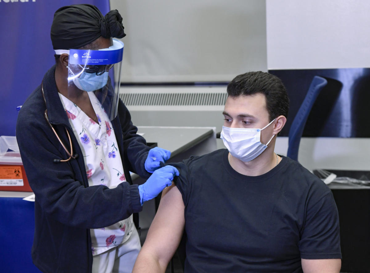
Vaccines against severe acute respiratory syndrome coronavirus 2 (SARS-CoV-2) are the most important countermeasure to fight the coronavirus 2019 (Covid-19) pandemic. From December 2020 through March 2021, the European Medicines Agency approved four vaccines on the basis of randomized, blinded, controlled trials: two messenger RNA–based vaccines — BNT162b2 (Pfizer–BioNTech) and mRNA-1273 (Moderna) — that encode the spike protein antigen of SARS-CoV-2, encapsulated in lipid nanoparticles; ChAdOx1 nCov-19 (AstraZeneca), a recombinant chimpanzee adenoviral vector encoding the spike glycoprotein of SARS-CoV-2; and Ad26.COV2.S (Johnson & Johnson/Janssen), a recombinant adenovirus type 26 vector encoding SARS-CoV-2 spike glycoprotein.

As of April 7, 2021, more than 82 million vaccine doses had been administered in the European Union; in Germany, approximately one quarter of vaccine recipients had received the ChAdOx1 nCov-19 vaccine.1 Beginning in late February 2021, several cases of unusual thrombotic events in combination with thrombocytopenia were observed in patients after vaccination with ChAdOx1 nCov-19.

Index Case

A previously healthy 49-year-old health care worker received her first dose of ChAdOx1 nCov-19 in mid-February 2021 (day 0). Over the next few days, she reported having minor symptoms (fatigue, myalgia, and headache). Beginning on day 5, she reported having chills, fever, nausea, and epigastric discomfort; she was admitted to a local hospital on day 10.

Table 1.

Laboratory Characteristics of the Index Patient.

Laboratory results are shown in Table 1. The platelet count was 18,000 per cubic millimeter, and the d-dimer level was 35 mg per liter (reference value, <0.5). The results of other blood tests were normal except for γ-glutamyltransferase and C-reactive protein levels, which were elevated. SARS-CoV-2 reverse-transcriptase–polymerase-chain-reaction assay of a nasopharyngeal swab was negative.

Computed tomography (CT) showed portal-vein thrombosis and peripheral pulmonary emboli. The patient received a platelet concentrate and was transferred to a tertiary hospital. On arrival, she had epigastric discomfort and nausea but was otherwise in good condition (blood pressure, 125/88 mm Hg; heart rate, 65 beats per minute; temperature, 36.5°C). The physical examination was unremarkable except for moderate epigastric pain on palpation. She received intravenous antibiotics, analgesia, and one 4000-unit dose of low-molecular-weight heparin (enoxaparin), given subcutaneously.

The following day, the platelet count and fibrinogen level remained low, and the d-dimer and aminotransferase levels increased. The abdominal pain worsened, and repeat CT imaging showed progression of portal-vein thrombosis to include the splenic and upper mesenteric veins; in addition, small thrombi were visualized in the infrarenal aorta and both iliac arteries. Low-dose intravenous unfractionated heparin (500 IU per hour) was initiated but was stopped shortly thereafter because of a sudden onset of tachycardia and concern for gastrointestinal bleeding. The lactate level was 3.7 mmol per liter, and the patient was transferred to the intensive care unit. Repeat CT imaging revealed diffuse gastrointestinal bleeding with reduced perfusion of the intestinal wall and pancreas by splanchnic-vein thrombosis, along with ascites. She received red-cell and platelet transfusions, prothrombin complex concentrates, and recombinant factor VIIa but died on day 11. In addition to the diagnosed medical findings, autopsy revealed cerebral venous thrombosis.

Case Series

Table 2. Clinical and Laboratory Summary of 11 Patients with Available Clinical Information.

By March 15, 2021, an additional 10 patients for whom clinical data were available were found to have one or more thrombotic complications beginning 5 to 16 days after vaccination with ChAdOx1 nCov-19. Characteristics of all 11 patients (including the index case) are presented in Table 2. Thrombotic events included cerebral venous thrombosis (in 9 patients), splanchnic-vein thrombosis (in 3 patients), pulmonary embolism (in 3 patients), and other types of thrombi (in 4 patients); 5 of 10 patients had more than one thrombotic event. Included in this analysis is one patient (Patient 11) who presented with fatal cerebral hemorrhage. The results of brain neuropathological analysis were pending at the time of this report, and cerebral venous thrombosis had not been ruled out; postmortem serum was available for testing for platelet-activating antibodies.

Among these patients, the median age was 36 years (range, 22 to 49); 9 of 11 were women. All the patients presented with concomitant thrombocytopenia (median nadir of platelet count, approximately 20,000 per cubic millimeter; range, 9000 to 107,000). One patient had preexisting von Willebrand disease, anticardiolipin antibodies, and factor V Leiden. None of the patients had received heparin before the onset of symptoms or the diagnosis of thrombosis. Given the striking clinical resemblance of this disorder to autoimmune heparin-induced thrombocytopenia (a prothrombotic thrombocytopenic disorder that can be triggered by heparin and certain other anions and that features heparin-independent platelet-activating properties), serum obtained from 4 of the 11 patients was referred for immediate investigation of platelet-activating antibodies directed against platelet factor 4 (PF4)–heparin. After characterizing the antibodies in serum obtained from Patients 1 through 4, we subsequently obtained serum from 5 of the 7 remaining patients. In addition, our reference laboratory received further serum samples from patients who were suspected of having prothrombotic thrombocytopenia related to ChAdOx1 nCov-19 vaccination. (No detailed clinical information regarding these patients was available at the time of this report.)

Methods

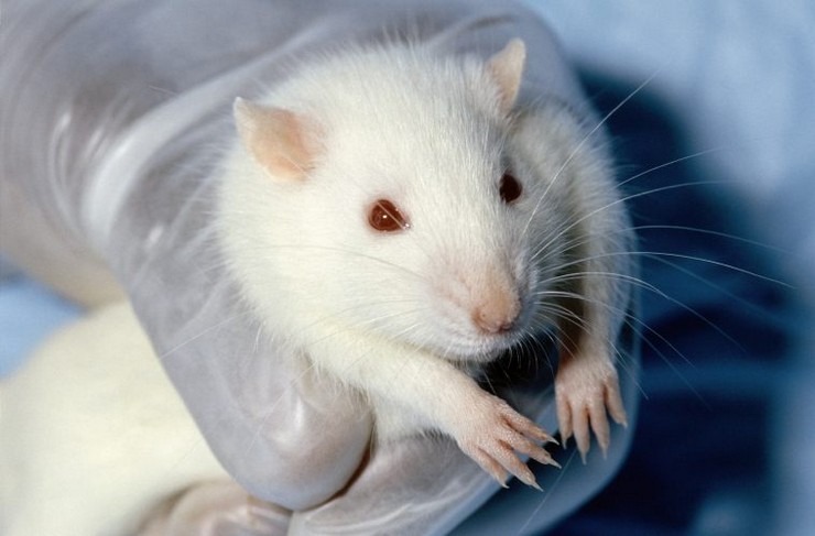
We purified platelets from whole blood (obtained from healthy volunteers) that had undergone anticoagulation with adenine citrate dextrose solution A. None of the volunteers had been taking antiplatelet drugs or had been vaccinated in the previous 10 days. We prepared platelets using methods that have been described previously.2,3 In a subgroup of experiments, platelets were preincubated in buffer with ChAdOx1 nCov-19 (1:2000 dilution) and washed before use. Washed platelets (75 microliters) were incubated with either buffer, a low-molecular-weight heparin (reviparin [Abbott]), or PF4 (Chromatec) in either the presence or absence of the FcγIIa receptor–blocking antibody IV.3. In some experiments, unfractionated heparin (100 IU per milliliter) was added to inhibit PF4-dependent reactions, or ChAdOx1 nCov-19 (1:50 dilution) was added per well. Serum was coincubated with PF4 and platelets in the presence of immune globulin (Privigen IVIG [CSL Behring]) at a concentration of 10 mg per milliliter. After establishing assay conditions using serum from the initial four patients, we investigated another 24 serum samples that tested positive on immunoassay to validate our findings. We refer to this modified platelet-activation test as the PF4-enhanced platelet-activation test.

To measure direct antibody binding, we used two immunoassays, a PF4–heparin enzyme-linked immunosorbent assay (ELISA) and a PF4 ELISA, with antibody binding measured by a secondary antihuman IgG, as described previously.4 In addition, antibodies from two serum samples were affinity purified by immobilized PF4–heparin and immobilized PF4, and the purified antibodies were tested in the assays. (Details about this method are provided in the Supplementary Appendix, available with the full text of this article at NEJM.org.)

We defined reactivity on ELISA according to the optical-density units as strong (≥2.00), intermediate (1.00 to 1.99), or weak (0.50 to 0.99). On the PF4-enhanced platelet-activation test, reactivity was graded according to the time that had elapsed until platelet aggregation,5 with shorter reaction times indicating stronger platelet activation (strong activation, 1 to 5 minutes; intermediate activation, >5 to 15 minutes; and weak activation, >15 to 30 minutes).

Results

All 11 patients in the initial analysis had moderate-to-severe thrombocytopenia and unusual thrombosis, particularly cerebral venous thrombosis and splanchnic-vein thrombosis (Table 2). We also found evidence of disseminated intravascular coagulation in 5 of the patients on the basis of the combination of greatly elevated d-dimer levels (>10.0 mg per liter) and one or more abnormalities in the international normalized ratio, partial thromboplastin time, or fibrinogen level. (Of the 6 patients with available fibrinogen levels, 4 had hypofibrinogenemia.)

Although evaluating the outcomes of different management strategies was not the goal of our study, we noted with interest the clinical course of Patient 2, who presented with pulmonary embolism and mild thrombocytopenia (platelet count, 107,000 per cubic millimeter), without disseminated intravascular coagulation. This patient received therapeutic-dose low-molecular-weight heparin for 3 days, with clinical improvement and an increase in the platelet count to 132,000; at that time, a positive result on PF4–heparin ELISA was obtained, and the patient was switched to oral apixaban, with continued clinical and laboratory recovery.

Figure 1. Reactivity of Patient Serum on Platelet-Activation Assays and Immunoassays.

Table 2 also shows results of the PF4–heparin ELISA, including for the first 4 patients in whom detailed laboratory studies were performed. Serum obtained from these patients showed strong reactivity on PF4–heparin ELISA, with optical densities of more than 3.00 units (reference value, <0.50); all reactivity reactions were inhibited to less than 0.50 units by the addition of heparin (100 IU per milliliter). Figure 1 shows the serologic profile of the 4 initial patients, as assessed by means of the platelet-activation assay. Three of the four serum samples showed weak-to-moderate reactivity at buffer control, which was inhibited by low-molecular-weight heparin. In three of the samples, PF4 (10 μg per milliliter) greatly enhanced reactivity; serum from Patient 2 subsequently showed strong platelet activation in the presence of PF4 when retested along with platelets from other volunteers. All reactions were blocked by monoclonal antibody IV.3 and immune globulin at a dose of 10 mg per milliliter, which indicated that platelet activation had occurred through platelet Fcγ receptors (Figure 1A). None of the controls showed platelet activation (data not shown).

Platelet activation was enhanced when platelets were pelleted from platelet-rich plasma, resuspended in washing buffer, preincubated (1:2000) with ChAdOx1 nCov-19, centrifuged, and resuspended in the final suspension buffer or when they were coincubated in the suspension buffer with ChAdOx1 nCov-19 (1:50). The monoclonal antibody IV.3 blocked PF4-dependent platelet activation in all 7 samples that were tested.

Figure 1B shows the results of platelet activation in serum samples obtained from 24 patients with clinically suspected vaccine-induced immune thrombotic thrombocytopenia who tested positive on the screening PF4–heparin ELISA. Whereas approximately half the serum samples (13 of 24) showed platelet activation at buffer control, most samples (19 of 24) were inhibited by low-molecular-weight heparin; almost all samples (22 of 24) showed platelet activation by the addition of PF4. All but one serum sample was inhibited by a high dose of heparin.

Figure 1C shows strong reactivity of the serum samples obtained from all 28 patients (including Patients 1, 2, 3, 4, 5, 8, 9, 10, and 11) in results on both PF4–heparin and PF4 ELISA, with inhibition by high heparin doses. Antibodies that were affinity purified with the use of either immobilized PF4 or immobilized PF4–heparin showed the same reactivity pattern as the original serum — in other words, they strongly activated platelets in the presence of 10 μg per milliliter of PF4, an effect that was completely inhibited by a high concentration of heparin.

Discussion

The clinical picture of moderate-to-severe thrombocytopenia and thrombotic complications at unusual sites beginning approximately 1 to 2 weeks after vaccination against SARS-CoV-2 with ChAdOx1 nCov-19 suggests a disorder that clinically resembles severe heparin-induced thrombocytopenia, a well-known prothrombotic disorder caused by platelet-activating antibodies that recognize multimolecular complexes between cationic PF4 and anionic heparin.6 However, unlike the usual situation in heparin-induced thrombocytopenia, these vaccinated patients did not receive any heparin to explain the subsequent occurrence of thrombosis and thrombocytopenia.

In recent years, it has been recognized that triggers other than heparin can cause a prothrombotic disorder that strongly resembles heparin-induced thrombocytopenia on both clinical and serologic grounds, including certain polyanionic drugs (e.g., pentosan polysulfate,7 antiangiogenic agent PI-88,8 and hypersulfated chondroitin sulfate8). Such a prothrombotic syndrome has also been observed in the absence of preceding exposure to any polyanionic medication, such as after both viral and bacterial infections9,10 and knee-replacement surgery.11,12 These various clinical scenarios with apparent nonpharmacologic triggers have been classified under the term autoimmune heparin-induced thrombocytopenia.13 Unlike patients with classic heparin-induced thrombocytopenia, patients with autoimmune heparin-induced thrombocytopenia have unusually severe thrombocytopenia, an increased frequency of disseminated intravascular coagulation, and atypical thrombotic events. Serum from these patients strongly activate platelets in the presence of heparin (0.1 to 1.0 IU per milliliter) but also in the absence of heparin (heparin-independent platelet activation). When these unusual antibodies are observed in patients who have thrombocytopenia without preceding heparin exposure, the term “spontaneous” heparin-induced thrombocytopenia syndrome13,14 has been used. Sometimes, patients in whom heparin-induced thrombocytopenia develops after exposure to heparin present with atypical clinical features, such as an onset of thrombocytopenia beginning several days after stopping heparin (delayed-onset heparin-induced thrombocytopenia15,16) or thrombocytopenia that persists for several weeks despite the discontinuation of heparin (persisting or refractory heparin-induced thrombocytopenia17,18). Serum from these patients also shows the phenomenon of heparin-independent platelet-activating properties.

These clinical features that resemble those of autoimmune heparin-induced thrombocytopenia were observed in the patients with vaccine-induced immune thrombotic thrombocytopenia. The serum usually showed strong reactivity on the PF4–heparin ELISA. Moreover, serum showed variable degrees of platelet activation in the presence of buffer that was in most cases greatly enhanced in the presence of PF4 (Figure 1A and 1B). More strikingly, most serum showed inhibition, rather than increased activation, in the presence of low-dose low-molecular-weight heparin (0.2 U per milliliter of anti–factor Xa). In addition, antibodies from two patients, which were affinity purified on either immobilized PF4 or immobilized PF4–heparin, strongly activated platelets but only in the presence of PF4. Enhancement of platelet activation by PF4 is also a feature of heparin-induced thrombocytopenia19,20 and has been used to enhance detection of platelet-activating antibodies in diagnostic testing for this adverse drug reaction.21 Whether these antibodies are autoantibodies against PF4 induced by the strong inflammatory stimulus of vaccination or antibodies induced by the vaccine that cross-react with PF4 and platelets requires further study.

Although we found enhanced reactivity of patient serum with platelets in the presence of ChAdOx1 nCov-19, this is likely to be an in vitro artifact. It is well known that adenovirus binds to platelets22 and causes platelet activation.22,23 Furthermore, the amount of adenovirus in a 500-microliter vaccine injection administered 1 or 2 weeks earlier would seem unlikely to contribute to subsequent platelet activation observed in these patients. However, interactions between the vaccine and platelets or between the vaccine and PF4 could play a role in pathogenesis. One possible trigger of these PF4-reactive antibodies could be free DNA in the vaccine. We have previously shown that DNA and RNA form multimolecular complexes with PF4, which bind antibodies from patients with heparin-induced thrombocytopenia and also induce antibodies against PF4–heparin in a murine model.24 Unfortunately, other Covid-19 vaccines were not available to us for testing.

Our findings have several important clinical implications. First, clinicians should be aware that in some patients, venous or arterial thrombosis can develop at unusual sites such as the brain or abdomen, which becomes clinically apparent approximately 5 to 20 days after vaccination. If such a reaction is accompanied by thrombocytopenia, it can represent an adverse effect of the preceding Covid-19 vaccination. To date, this reaction has been reported only with the ChAdOx1 nCov-19 vaccine, which has been used in approximately 25% of vaccine recipients in Germany and in 30% of those in Austria.

Second, ELISA to detect PF4–heparin antibodies in patients with heparin-induced thrombocytopenia is widely available and can be used to investigate patients for potential postvaccination thrombocytopenia or thrombosis associated with antibodies against PF4.25 A strongly positive ELISA result that is obtained in a patient who has not been recently exposed to heparin would be a striking abnormality.

Third, we have shown that these antibodies recognize PF4 and that the addition of PF4 greatly enhances their detectability in a platelet-activation assay. Since vaccination of millions of persons will be complicated by a background of thrombotic events unrelated to vaccination, a PF4-dependent ELISA or a PF4-enhanced platelet-activation assay may be used to confirm the diagnosis of vaccine-induced immune thrombotic thrombocytopenia through this novel mechanism of postvaccination formation of platelet-activating antibodies against PF4. Although treatment decisions such as administering intravenous immune globulin and starting anticoagulation do not need to await laboratory diagnosis, detection of these unusual platelet-activating antibodies will be highly relevant for case identification and future risk–benefit assessment of this and other vaccines.

Figure 2.


Potential Diagnostic and Therapeutic Strategies for Management of Suspected Vaccine-Induced Immune Thrombotic Thrombocytopenia.

Figure 2 shows a potential diagnostic and therapeutic strategy for managing this novel prothrombotic thrombocytopenic disorder. One consideration is to administer high-dose intravenous immune globulin to inhibit Fcγ receptor–mediated platelet activation. This recommendation parallels emerging experience in the treatment of severe autoimmune heparin-induced thrombocytopenia in which high-dose intravenous immune globulin has resulted in rapid increases in platelet count and de-escalation of hypercoagulability.12,26 We found that the addition of immune globulin in doses that are readily achieved clinically was effective in inhibiting platelet activation by patients’ antibodies. Clinician reluctance to start anticoagulation may be tempered by administering high-dose intravenous immune globulin to raise the platelet count, especially when a patient presents with severe thrombocytopenia and thrombosis, such as cerebral venous thrombosis.

Given the parallels with autoimmune heparin-induced thrombocytopenia, anticoagulant options should include nonheparin anticoagulants used for the management of heparin-induced thrombocytopenia,27 unless a functional test has excluded heparin-dependent enhancement of platelet activation. Finally, we suggest naming this novel entity vaccine-induced immune thrombotic thrombocytopenia (VITT) to avoid confusion with heparin-induced thrombocytopenia.

Supported by a grant (374031971–TRR 240) from the German Research Foundation.

Disclosure forms provided by the authors are available with the full text of this article at NEJM.org.

Drs. Greinacher and Thiele contributed equally to this article.

This article was published on April 9, 2021, at NEJM.org.

We thank the colleagues who provided serum samples for this study, including Dr. Michael Hirschl of Landesklinikum Zwettl, Austria; Dr. Johannes Thaler of the Medical University of Vienna; Dr. Brigitte Keller-Stanislawski, Dr. Dirk Mentzer, and Prof. Dr. Klaus Cichutek of the Paul-Ehrlich-Institut, Langen, Germany; and Prof. Dr. Hans-Georg Bone, Dr. Juliane Alfes, and Dr. Hans-Christian Atzpodien of Klinikum Vest, Recklinghausen; technologists Ulrike Strobel, Carmen Freyer, Katrin Stein, Ines Warnig, and Ricarda Raschke of Transfusion Medicine Greifswald; and the members of the council of the Gesellschaft für Thrombose und Hämostaseforschung (Prof. Dr. Johannes Oldenburg, Dr. Robert Klamroth, Prof. Dr. Florian Langer, and Prof. Dr. Bernd Pötzsch) for their support.

Author Affiliations

From Institut für Immunologie und Transfusionsmedizin, Universitätsmedizin Greifswald, Greifswald (A.G., T.T.), and the Division of Safety of Medicinal Products and Medical Devices, Paul-Ehrlich-Institut (Federal Institute for Vaccines and Biomedicines), Langen (K.W.) — both in Germany; the Departments of Pathology and Molecular Medicine and of Medicine, McMaster University, Hamilton, ON, Canada (T.E.W.); and the Department of Medicine I, Division of Hematology and Hemostaseology, Medical University of Vienna, Vienna (P.A.K., S.E.).

Address reprint requests to Dr. Greinacher at Institut für Immunologie und Transfusionsmedizin, Abteilung Transfusionsmedizin, Sauerbruchstrasse, 17487 Greifswald, Germany.

https://www.nejm.org/doi/10.1056/NEJMoa2104840

NEJ means New England Journal of Medicine

Ραββίνος κατά των Χαζαρο-Ιουδαίων. Καμιά σχέση των Εβραίων με τους Χαζάρ.

Παρασκευή, 14 Μαΐου 2021

ΤΙ ΛΕΕΙ Ο ΡΑΒΒΙΝΟΣ ΡΕ ΠΑΙΔΙΑ…;; ΕΧΩ ΠΑΘΕΙ ΚΟΚΟΜΠΛΟΚΟ!

Ραββίνος Yossef Ben Porat:

“Τα αίτια του μεγάλου διωγμού”

 Βίντεο που θα μείνει στην Ιστορία!

(αναρτήθηκε πριν από 2 ώρες)

                [όχι πως εμείς δεν τα ξέραμε..]


https://www.youtube.com/watch?v=HQdZfzcTWyk


Ασφαλώς και το έκοψαν οι φασίστες του Youtube.