Μας δολοφονούν ύπουλα ξυπνήστε επιτέλους!

Η Δουλο-Κυβέρνηση σε υποχρεώνει για μάσκα ΚΝ-95 όταν στη Γαλλία ανεστάλη η χρήση τους!!!

Ενώ στην Ελλάδα την κάνουμε υποχρεωτική για σούπερ μάρκετ και Μέσα Μαζικής Μεταφοράς στη Γαλλία έχει ανασταλεί η χρήση της γιατί η εθνική υπηρεσία ασφάλειας υγείας της Γαλλίας (ANSES) αποφάσισε ότι οι μάσκες FFP2 περιέχουν γραφένιο και δεν πρέπει πλέον να χρησιμοποιούνται, λόγω έλλειψης δεδομένων σχετικά με την τοξικότητα τους.

Υποχρεωτική θα είναι πλέον στην Ελλάδα η χρήση της μάσκας ΚΝ-95, για είσοδο των πολιτών σούπερ μάρκετ και στα Μέσα Μαζικής Μεταφοράς. Αυτό ανακοίνωσε ο υπουργός Υγείας, Θ. Πλεύρης, τη συγκεκριμένη μάσκα θα την έχουμε πλέον όλοι μαζί, ειδικά όσοι πηγαίνουν για ψώνια ή χρησιμοποιούν μέσα μεταφοράς.

«Η ANSES συνιστά στις δημόσιες αρχές να δίνουν προτεραιότητα στην εμπορία ή τη διάθεση μασκών χωρίς γραφένιο»

Η χρήση μασκών FFP2 που περιέχουν γραφένιο, έχει ήδη ανασταλεί προσωρινά στη Γαλλία, πρέπει να αποφευχθεί προληπτικά λόγω έλλειψης στοιχείων για την τοξικότητα αυτού του συνθετικού υλικού, έτσι έκρινε η υπηρεσία υγειονομικής ασφάλειας Anses. και ανακοίνωσε μέσο δελτίο τύπου μετά από έκθεση εμπειρογνωμόνων που ζήτησε η Γενική Διεύθυνση Υγείας.

Τον Απρίλιο, ο Καναδάς ήταν η πρώτη χώρα που τόνισε τους πιθανούς κινδύνους των μασκών FFP2 (ή KN95) για τον ίδιο λόγο, επειδή περιέχουν γραφένιο. Είχε αφαιρέσει προληπτικά τις μάσκες γραφενίου που κατασκεύαζε η κινεζική εταιρεία Shandong Shengquan New Materials, που τότε υποπτευόταν ότι προκαλούσε προβλήματα στους πνεύμονες. Μετά από ανάλυση, ο Καναδάς ενέκρινε τελικά ξανά αυτές τις μάσκες τον Ιούλιο, θεωρώντας ότι δεν παρουσίαζαν αποδεδειγμένο κίνδυνο.

Η ANSES ωστόσο θεωρεί «αδύνατο να εκτιμηθεί ο κίνδυνος για την υγεία». Σημειώνει μάλιστα «η έλλειψη πληροφοριών για το γραφένιο που χρησιμοποιείται από τους κατασκευαστές, για την τοξικότητα αυτής της ουσίας, είναι αδύνατον να γνωρίζουμε τις επιπτώσεις ιδίως μακροπρόθεσμα». Γι’ αυτό θεωρεί προτιμότερο να αποφεύγεται η χρήση αυτού του είδους μάσκας.

anazitiseis.gr

ΔΙΑΒΑΣΤΕ ΕΔΩ 150 ΜΕΛΕΤΕΣ ΓΙΑ ΤΙΣ ΜΑΣΚΕΣ…brownstone.org

Μεγάλη βελτίωση από τη χρήση μαγνητικών πεδίων στη θεραπεία ασθενών με ρευματοειδή αρθρίτιδα.

Η χρήση μαγνητικών πεδίων στη θεραπεία ασθενών με ρευματοειδή αρθρίτιδα.  Ανασκόπηση της βιβλιογραφίας

Jolanta Zwolińska , 1, 2 Monika Gąsior , 2 Elżbieta Śnieżek , 1 και Andrzej Kwolek 1

Πληροφορίες συντάκτη Σημειώσεις άρθρου Σημειώσεις πνευματικών δικαιωμάτων και πληροφοριών άδειας Αποποίηση ευθύνης

Αυτό το άρθρο έχει αναφερθεί από άλλα άρθρα στο PMC.

Αφηρημένη

Εισαγωγή

Μαζί με την επιστημονική και τεχνική πρόοδο και την τεχνολογική ανάπτυξη, η φυσική ιατρική προσφέρει νέες θεραπευτικές επιλογές [  ]. Η θεραπεία με χρήση φυσικών παραγόντων απαιτεί υψηλά πρότυπα ασφάλειας και εμπειρίας παραπέμποντος γιατρού και θεραπευτή. Η φυσικοθεραπεία μπορεί να είναι μια σημαντική θεραπευτική μέθοδος που συμπληρώνει τη φαρμακευτική αγωγή ή την αποκατάσταση [  ,  ].

Η χρήση της θεραπείας με μαγνητικό πεδίο (MF) σχετίζεται με την εντατική ανάπτυξη της γνώσης σχετικά με την επίδραση αυτού του πεδίου σε έναν ζωντανό οργανισμό σε επίπεδο κυττάρων και ιστών [  ]. Ωστόσο, η σύγχρονη γνώση του μηχανισμού της επίδρασης του MF στους ζωντανούς οργανισμούς εξακολουθεί να αφήνει πολλές αβεβαιότητες [  ]. Ο μηχανισμός των θεραπευτικών επιδράσεων του MF στο σύστημα δεν είναι πλήρως κατανοητός [  ]. Απαιτείται μια διεξοδική ανάλυση της φυσικής φύσης του MF καθώς και η πραγματοποίηση αντικειμενικών κλινικών δοκιμών για την αξιολόγηση της επίδρασης του πεδίου στο σύστημα για τη βελτιστοποίηση των αποτελεσμάτων της μαγνητικής θεραπείας [  , ]. Η ανάγκη επικύρωσης της ασφάλειας και της αποτελεσματικότητας της μαγνητοθεραπείας στην πορεία διαφόρων ασθενειών και συνδρόμων πηγάζει από τις κατευθυντήριες γραμμές της ιατρικής που βασίζεται σε στοιχεία (EBM) και αποτελεί πρόκληση για τους σημερινούς ερευνητές.

Το μαγνητικό πεδίο και η επίδρασή του στον οργανισμό

Η μαγνητική ενέργεια υπάρχει παντού στον κόσμο [  ]. Σύμφωνα με τον Heisenberg, είναι στοιχειώδης ενέργεια, από την οποία εξαρτάται η ζωή του οργανισμού [  ]. Το μαγνητικό πεδίο της Γης δείχνει την ένταση στην περιοχή των 30-70 μT, η οποία είναι ένας φυσικός διεγέρτης των φυσιολογικών διεργασιών σε έναν ζωντανό οργανισμό [  ]. Το φαινόμενο της αυξημένης επίπτωσης διαφόρων ασθενειών του ανθρώπινου οργανισμού εξηγείται από τη συνεχή μείωση της ισχύος του μαγνητικού πεδίου της Γης [  ,  ]. Παρατηρήσεις ανθρώπων που μένουν στο διάστημα για μεγάλο χρονικό διάστημα δείχνουν ότι η έλλειψη φυσικού μαγνητικού πεδίου προκαλεί αϋπνία, κόπωση και κατάθλιψη και αυξάνει τον κίνδυνο οστεοπόρωσης [  ].

Το MF προκαλεί κίνηση των ιόντων εντός του κυττάρου, η οποία έχει ως αποτέλεσμα την υπερπόλωση της κυτταρικής μεμβράνης. Αυτό επηρεάζει την επιτάχυνση του μεταβολισμού, ιδίως διεγείροντας τις ενεργειακές διεργασίες και αυξάνοντας τη χρήση οξυγόνου από το κύτταρο [  ,  ,  ]. Τέτοιες αλλαγές είναι διεγερτικά για τις διαδικασίες πολλαπλασιασμού ή απόπτωσης [  ]. Το MF έχει ευεργετική επίδραση στους αναγεννητικούς μηχανισμούς που εμφανίζονται στον ιστό και διεγείρει τη χυμική και ανοσοποιητική δραστηριότητα σε ολόκληρο τον οργανισμό [  , ]. Ενισχύει τις ορμονικές και ενζυμικές αντιδράσεις. Το MF δρα επίσης στα οστά και στον συνδετικό ιστό. Τα ηλεκτρικά ρεύματα που προκαλούνται στους ιστούς μπορεί να επηρεάσουν τις πιεζοηλεκτρικές ιδιότητες των υλικών, προκαλώντας μηχανική παραμόρφωση, η οποία διεγείρει το σχηματισμό τύλου σε περίπτωση απόφραξης στην επούλωση των οστών [  ]. Η εφαρμογή του MF για θεραπευτικούς σκοπούς βασίζεται σε μελέτες που επιβεβαίωσαν την αντιφλεγμονώδη, αντιοιδηματική, αγγειοδιασταλτική και αγγειογενετική δράση [  ,  ]. Μαγνητοθεραπεία έχει επίσης ένα αναλγητικό αποτέλεσμα, επηρεάζει την ένταση των μυών, και διεγείρει την αναγέννηση των ιστών [  ,  – ]. Έχει μια κατευναστική επίδραση, επηρεάζει την ευεξία και έχει κατά προτίμηση επιρροές στην ανθρώπινη ψυχή [  ,  ].

Η αποτελεσματική εφαρμογή της μαγνητοθεραπείας απαιτεί εφαρμογή των συνιστώμενων παραμέτρων πεδίου και τήρηση των οδηγιών της μεθοδολογίας θεραπείας [  ,  ]. Οι θεραπείες μαγνητοθεραπείας απαιτούν διαφορετικούς τύπους γεννητριών πεδίου, την ένταση και την πορεία του πεδίου καθώς και διαφορετικούς τρόπους εφαρμογής [  ,  ,  ]. Αυτές οι τιμές επιλέγονται μεμονωμένα ανάλογα με τη γενική κατάσταση του ασθενούς και το στάδιο της νόσου [  ]. Το μαγνητικό πεδίο επιτρέπει τη βαθιά διείσδυση του ιστού λόγω της διεισδυτικής ισχύος των διαφόρων δομών, συμπεριλαμβανομένων των γύψινων εκμαγείων [  ,  ].

Η μαγνητοθεραπεία μπορεί να εφαρμοστεί με ασφάλεια για μεγαλύτερες σειρές (10–15 θεραπείες ή περισσότερες). Η αρχική θεραπεία πραγματοποιείται καθημερινά, στη συνέχεια κάθε δεύτερη μέρα και η τελευταία θεραπεία έως 2 φορές την εβδομάδα [  ]. Το αναλγητικό αποτέλεσμα της μαγνητοθεραπείας αυξάνεται με τον αριθμό των διαδικασιών που εκτελούνται [  ]. Οι θεραπείες διάρκειας δεκαπέντε λεπτών μπορούν μερικές φορές να παραταθούν σε 60 λεπτά, ενώ δεν συνιστάται η χρήση θεραπειών που διαρκούν λιγότερο από 5 λεπτά [  ]. Οι περισσότερες συνιστώμενες θεραπείες διαρκούν 30 λεπτά [  ]. Δεν συνιστώνται θεραπείες αργά το βράδυ [ ]. Η θεραπεία με ένα παλλόμενο ηλεκτρομαγνητικό πεδίο (PEMF) συχνά συνδυάζεται με άλλες φυσικές και λουτροθεραπευτικές διαδικασίες, οι οποίες χρησιμοποιούνται μαζί βελτιώνουν το θεραπευτικό αποτέλεσμα [  ,  ,  ]. Η ουσιαστική προετοιμασία των φυσιοθεραπευτών για την εκτέλεση διαδικασιών καθώς και η συμβατότητα των τιμών που δηλώνονται από έναν κατασκευαστή με την πραγματική κατάσταση είναι παράγοντες που καθορίζουν τα θεραπευτικά αποτελέσματα [  ].

Οι θεραπείες που χρησιμοποιούν μόνιμους μαγνήτες όπου το μαγνητικό πεδίο δημιουργείται από έναν μαγνήτη που συνδέεται με ρούχα, μαξιλάρια, κοσμήματα, παπούτσια κ.λπ. χρησιμοποιούνται επίσης στη μαγνητοθεραπεία [  ]. Οι μόνιμοι μαγνήτες είναι εύκολοι στη χρήση, φθηνοί και ευρέως διαθέσιμοι. Η χρήση τέτοιων μαγνητών είναι μια μορφή θεραπείας του χρόνιου πόνου που σχετίζεται με δυσλειτουργία του μυοσκελετικού συστήματος και φλεγμονώδεις διαταραχές συμπεριλαμβανομένης της ρευματοειδούς αρθρίτιδας (RA) [  ]. Η τρέχουσα κατάσταση γνώσης δεν επαρκεί για να επιβεβαιώσει ξεκάθαρα τη θεραπευτική αποτελεσματικότητα των στατικών μαγνητικών πεδίων [ ]. Δεν χαρακτηρίζονται όλες οι κλινικές δοκιμές για τη δράση των μόνιμων μαγνητών που έχουν πραγματοποιηθεί μέχρι τώρα από υψηλή μεθοδολογική ποιότητα και τα αποτελέσματα αυτών των δοκιμών δεν μπορούν να παρέχουν επιστημονικά στοιχεία για την αποτελεσματικότητα μιας τέτοιας θεραπείας σε όλες τις περιπτώσεις [  ].

Ασφάλεια μαγνητοθεραπείας

Το μαγνητικό πεδίο δεν προκαλεί θερμική επίδραση στον ιστό και δεν γίνεται άμεσα αντιληπτό από το ανθρώπινο σώμα [  ,  ,  ]. Η μεγαλύτερη ένταση αυτού του πεδίου αναφέρεται από τα άτομα ως διάφορα είδη συναισθημάτων, όπως μυρμήγκιασμα και δόνηση [  ]. Δεν σημειώθηκαν σημαντικές παρενέργειες κατά τη χρήση τέτοιων πεδίων [  ]. Στην περίπτωση υπερευαισθησίας, είναι πιθανά νευρασθενικά και φυτικά συμπτώματα, δερματολογικά συμπτώματα (ερυθρότητα, μυρμήγκιασμα, κάψιμο), θολή όραση, αυξημένη ευαισθησία σε χημικά ερεθίσματα και άλλα συμπτώματα [ ]. Ο Διεθνής Οργανισμός Έρευνας για τον Καρκίνο ταξινόμησε τα μαγνητικά πεδία EMF-LF χαμηλής συχνότητας (ηλεκτρομαγνητικά πεδία χαμηλής συχνότητας) ως πιθανό καρκινογόνο παράγοντα [  ]. Τα στατικά μαγνητικά πεδία θεωρούνται ασφαλή ακόμη και για πολύ υψηλή μαγνητική επαγωγή, ενώ οι επιβλαβείς επιπτώσεις των ηλεκτρομαγνητικών πεδίων στο ανθρώπινο σώμα με μακροχρόνια έκθεση σε αυτά έχουν αποδειχθεί [  ,  ,  ]. Είναι σημαντικό να ληφθούν υπόψη οι πιθανοί κίνδυνοι και η συμμόρφωση με τους ισχύοντες κανόνες για τις διαδικασίες [  ].

Ρευματοειδής αρθρίτιδα – επιπτώσεις στην αποτελεσματικότητα και την ποιότητα ζωής

Η συχνότητα της ρευματοειδούς αρθρίτιδας στον κόσμο κυμαίνεται από 0,3 έως 1,5% του πληθυσμού, ενώ στην Ευρώπη επηρεάζει το 0,8% του ενήλικου πληθυσμού [  ]. Υπολογίζεται ότι στην Πολωνία, περίπου 400.000 άνθρωποι πάσχουν από φλεγμονώδεις ασθένειες των αρθρώσεων, συμπεριλαμβανομένων 135.000–157.000 που πάσχουν από ΡΑ. Παρατηρείται συνεχής αύξηση του αριθμού των κρουσμάτων [  ]. Η ΡΑ συνήθως επηρεάζει άτομα ηλικίας 30-50 ετών. Οι γυναίκες υποφέρουν 2-3 φορές πιο συχνά. Σε σύγκριση με τον γενικό πληθυσμό, οι ασθενείς με ΡΑ ζουν 7-10 χρόνια μικρότερη ζωή [  ,  ]. Το οικονομικό κόστος που προκύπτει από αυτή την ασθένεια σχετίζεται κυρίως με την απώλεια της ικανότητας για εργασία και την ανάγκη παροχής φροντίδας σε περίπτωση απώλειας της αποτελεσματικότητας [ ].

Η ρευματοειδής αρθρίτιδα είναι μια χρόνια και προοδευτική πάθηση. Η συνεχιζόμενη φλεγμονώδης διαδικασία οδηγεί σε παραμόρφωση των αρθρώσεων, εξασθένηση της λειτουργίας τους, επιπλοκές στα εσωτερικά όργανα και ψυχικές αλλαγές. Η ασθένεια οδηγεί σε σημαντική μείωση της ποιότητας ζωής, αναπηρία και πρόωρο θάνατο [  ]. Τα συμπτώματα της κατάθλιψης εμφανίζονται στο 13-25% των ασθενών με ΡΑ, όταν ο ασθενής συνειδητοποιεί τη δυσμενή πρόγνωση σε αυτή τη νόσο [  ]. Τα χαρακτηριστικά κλινικά χαρακτηριστικά της ΡΑ περιλαμβάνουν πόνο – συχνά χρόνιο – οίδημα, περιορισμό της κίνησης, δυσκαμψία και φλεγμονή που εμφανίζεται σε πολλαπλές αρθρώσεις [  ,  , ]. Ο πόνος είναι ο πιο σημαντικός παράγοντας που καθορίζει το επίπεδο αναπηρίας του ασθενούς [  ]. Ο πόνος στις αρθρώσεις προκαλεί το αντανακλαστικό τάσης των μυών, το οποίο προκαλεί συμπίεση των κατεστραμμένων αρθρώσεων και επιδεινώνει τον πόνο. Δημιουργείται ο μηχανισμός του λεγόμενου φαύλου κύκλου του πόνου [  ,  ]. Η σωματική δραστηριότητα του ασθενούς είναι περιορισμένη, παρατηρείται σταδιακή απώλεια μυϊκού και οστικού ιστού, διαταραχές ύπνου και γενική κόπωση. Ο ασθενής εξαρτάται περισσότερο από το περιβάλλον [  ,  ,  ].

Η θεραπεία ασθενών με ΡΑ είναι πολλαπλής δράσης, που περιλαμβάνει φαρμακολογικές, χειρουργικές και αποκαταστατικές προσεγγίσεις. Το εύρος της θεραπείας και της αποκατάστασης εξαρτάται από τη λειτουργική κατάσταση του ασθενούς, την ηλικία, τη διάρκεια της νόσου και την έκταση της βλάβης στο μυοσκελετικό σύστημα [  ,  ,  ]. Η αποκατάσταση σε κάθε στάδιο επιβραδύνει την εξέλιξη της διαδικασίας της νόσου και βελτιώνει την ποιότητα ζωής των ασθενών με ΡΑ [  ,  ,  ]. Οι εργαστηριακές εξετάσεις καθιστούν δυνατή την αξιολόγηση της αποτελεσματικότητας της θεραπείας και την πιθανή τροποποίησή της [  , ]. Η αποκατάσταση των ασθενών με ΡΑ είναι μια πολύπλοκη διαδικασία που διεξάγεται από τη στιγμή της διάγνωσης έως το τέλος της ζωής [  ].

Ως μέρος της ολοκληρωμένης αποκατάστασης, εφαρμόζονται φυσικοί παράγοντες που έχουν αναλγητική δράση, μειώνουν την πρωινή δυσκαμψία, βελτιώνουν τη λειτουργική κατάσταση του ασθενούς και περιορίζουν τη χρήση αναλγητικών φαρμάκων [  ,  ,  ]. Τα πρώτα αισθητά αποτελέσματα εμφανίζονται μετά από μια σειρά θεραπειών. Η ποιότητα των αποτελεσμάτων επηρεάζεται θετικά από τη συνέχιση των θεραπειών στο spa ( lat sanus per aqua ), όπου υπάρχει η δυνατότητα εμπλουτισμού της ολοκληρωμένης φυσικοθεραπείας για λουτροθεραπεία. Μια τέτοια προσέγγιση αυξάνει το αναλγητικό αποτέλεσμα και επεκτείνει την απόκριση [  ]. Η θεραπεία τόσο με μόνιμο όσο και με εξαιρετικά χαμηλής συχνότητας MF (ELF MF) εφαρμόζεται με επιτυχία μεταξύ των μεθόδων φυσικής θεραπείας.

Εφαρμογή μαγνητικού πεδίου σε ασθενείς με ρευματοειδή αρθρίτιδα

Το μαγνητικό πεδίο χαμηλής συχνότητας (LF-EMF) χρησιμοποιείται συνήθως στη θεραπεία ασθενών με ασθένειες του οστεοαρθρικού συστήματος συμπεριλαμβανομένης της ΡΑ [  ,  ]. Χρησιμοποιούνται τόσο θεραπείες μαγνητοθεραπείας (επαγωγή πάνω από 0,1 mT) όσο και μαγνητική διέγερση (επαγωγή κάτω από 0,1 mT) [  ]. Η θεραπεία μειώνει την αντιδραστικότητα του ανοσοποιητικού συστήματος σε περίπτωση φλεγμονωδών και εκφυλιστικών αλλαγών στις αρθρώσεις [  ]. Δίνει επίσης αναλγητικό και αντιοιδηματικό αποτέλεσμα [  ,  ]. Σύμφωνα με την Cieślińska-Świder [ ], μαγνητική ένταση 2 mT και συχνότητα 12 Hz χρησιμοποιούνται στην αρθρίτιδα. Ο συνιστώμενος χρόνος θεραπείας είναι από 15 έως 30 λεπτά και οι θεραπείες εκτελούνται 1-2 φορές την ημέρα για αρκετές εβδομάδες [  ].

Ο στόχος της έρευνας που πραγματοποιήθηκε από τους Kumar et al. [  ] επρόκειτο να αξιολογήσει την επίδραση των παλμικών ηλεκτρομαγνητικών πεδίων (PEMF) στην πειραματικά επαγόμενη φλεγμονή σε αρουραίους. Εφαρμόστηκε ένα ημιτονοειδές μονοπολικό μαγνητικό πεδίο με διαφορετικές παραμέτρους. Αξιολογήθηκε η δραστηριότητα των λυσοσωμικών ενζύμων, η διήθηση των φλεγμονωδών κυττάρων καθώς και η διόγκωση. Οι βέλτιστες παράμετροι πεδίου ήταν 4 mT, 5 Hz και χρόνος έκθεσης 90 λεπτά σε εκείνη την περίπτωση [  ]. Σε μελέτες από Dortch και Johnson [  ] αρουραίοι με πειραματικά επαγόμενη ρευματοειδή αρθρίτιδα υποβλήθηκαν καθημερινά σε μαγνητικό πεδίο AC με τις παραμέτρους 10 mT και 5-50 Hz. Έχει αποδειχθεί ότι το PEMF μπορεί να είναι ένας χρήσιμος θεραπευτικός παράγοντας στη θεραπεία χρόνιων φλεγμονωδών ασθενειών όπως η ρευματοειδής αρθρίτιδα [ ].

Shupak et al. [  ] διεξήγαγε μια διπλή-τυφλή, τυχαιοποιημένη δοκιμή με μια ομάδα ελέγχου που περιελάμβανε ασθενείς με ΡΑ και ινομυαλγία. Οι ομάδες μελέτης υποβλήθηκαν σε μαγνητοθεραπεία με δύο διαφορετικές τιμές της μέγιστης επαγωγής 200 μT και 400 μT. Οι μελέτες έδειξαν μια ελαφρά μείωση του επιπέδου του πόνου μετά τη χρήση της μαγνητοθεραπείας, αν και η αλλαγή δεν ήταν στατιστικά σημαντική. Οι συγγραφείς κατέληξαν στο συμπέρασμα ότι η μαγνητική θεραπεία παρουσιάζει μικρή αναλγητική δράση σε ασθενείς με ΡΑ [  ]. Οι μελέτες των Krawczyk-Wasielewska et al. [ ] διερεύνησε 50 ασθενείς με ΡΑ που υποβλήθηκαν σε διάφορους τύπους φυσιοθεραπείας (κρυοθεραπεία, υπερηχοθεραπεία, μαγνητικό πεδίο, θεραπεία με λέιζερ, διαδυναμική, μέθοδος TENS). Το πρήξιμο, το επίπεδο πόνου και η διάρκεια της πρωινής δυσκαμψίας αξιολογήθηκαν πριν και μετά τη θεραπεία. Δεν υπήρχαν συμπτώματα από το ουροποιητικό μετά την εφαρμογή οποιουδήποτε φυσικοθεραπευτικού παράγοντα. Οι συγγραφείς έδειξαν ότι το μαγνητικό πεδίο είχε ως αποτέλεσμα τη μείωση του πόνου στο 82% των ερωτηθέντων, ενώ το μέγεθος του αναλγητικού αποτελέσματος αξιολογήθηκε από τους συγγραφείς ως μέτριο. Παρατηρήθηκε μείωση του οιδήματος και μείωση της διάρκειας της πρωινής δυσκαμψίας. Η κρυοθεραπεία, η θεραπεία με λέιζερ, το TENS και η υπερηχοθεραπεία θεωρήθηκαν τα πιο αποτελεσματικά από τους συγγραφείς [  ].

Οι Greaves και Harlow [  ] αξιολόγησαν τη χρησιμότητα της εφαρμογής αδύναμων μαγνητών στην παρατήρηση των επιδράσεων εικονικού φαρμάκου στις μελέτες για τη μαγνητοθεραπεία. Χρησιμοποίησαν μαγνήτες των 180 mT ως ενεργούς μαγνήτες και μαγνήτες των 50 mT ως εικονικό φάρμακο. Οι συγγραφείς κατέληξαν στο συμπέρασμα ότι οι μαγνήτες των 50 mT μπορεί να μην εφαρμοστούν σε μια δοκιμή εικονικού φαρμάκου, καθώς στην υποκειμενική αξιολόγηση των ασθενών έχουν θεραπευτικό αποτέλεσμα [  ]. Οι μελέτες των Krawczyk-Wasielewska et al. [  ] έδειξε ότι η χρήση φυσικών παραγόντων σε άτομα με ρευματοειδή αρθρίτιδα (θερμοθεραπεία, υπερηχοθεραπεία, φωτοθεραπεία, ηλεκτροθεραπεία και μαγνητοθεραπεία) κατά τη διάρκεια παραμονής στο νοσοκομείο δύο εβδομάδων μείωσε τον πόνο και βελτίωσε τη λειτουργική κατάσταση και το επίπεδο συνολικής αποτελεσματικότητας σε όλους τους ασθενείς. [ ]. Kalmus et al. [  ] έδειξε ότι η χρήση μαγνητικών στρωμάτων που παράγουν ισχυρό μαγνητικό πεδίο με μέγιστη επαγωγή 64 mT μείωσε την κινητική δραστηριότητα κατά τη διάρκεια του ύπνου, γεγονός που συνέβαλε στη βελτίωση της ποιότητάς του. Υπήρξε μια στατιστικά σημαντική μείωση στο επίπεδο του πόνου καθώς και μια στατιστικά σημαντική μείωση στη δοσολογία των ΜΣΑΦ στην ομάδα που έλαβε θεραπεία με μαγνητοθεραπεία [  ].

Skalska-Izdebska et al. [  ] επιβεβαίωσε την αναλγητική και αντιοιδηματική δράση της μαγνητοθεραπείας σε ασθενείς με ΡΑ. Και τα 20 άτομα που υποβλήθηκαν σε μαγνητοθεραπεία ανέφεραν μείωση του περιαρθρικού οιδήματος και το 70% πέτυχε αναλγητικό αποτέλεσμα [  ]. Οι μελέτες των Walasik et al. [ ] αξιολόγησε την αποτελεσματικότητα ορισμένων σετ φυσικοθεραπείας που εκτελούνται στα χέρια ασθενών με ΡΑ σε εξωτερική βάση. Συγκρίθηκε η αποτελεσματικότητα δύο σετ φυσικοθεραπείας που χρησιμοποιούνται στην ομάδα Α (ηχοθεραπεία, θεραπεία με λέιζερ) και στην ομάδα Β (μαγνητική θεραπεία, κρυοθεραπεία). Και στις δύο ομάδες χρησιμοποιήθηκε το ίδιο κινησιοθεραπευτικό πρόγραμμα που περιλαμβάνει χειροκίνητες ασκήσεις χεριών. Διαπιστώθηκε ότι η εφαρμογή φυσικών διαδικασιών σε συνδυασμό με κινησιοθεραπεία περιορίζει αποτελεσματικά τον αριθμό των ευαίσθητων και διογκωμένων αρθρώσεων και μειώνει τον πόνο. Αποδείχθηκε ότι ένα σετ που αποτελείται από μαγνητοθεραπεία και κρυοθεραπεία είναι πιο αποτελεσματικό [  ]. Οι Chwieśko-Minarowska et al. [ ] συνέκρινε την επίδραση της διέγερσης με λέιζερ και του μαγνητικού πεδίου χαμηλής συχνότητας στη λειτουργία του χεριού και στην ποιότητα ζωής των ασθενών με ΡΑ. Χρησιμοποιήθηκε ένταση μαγνητικού πεδίου από 3 mT και συχνότητα 5 Hz, αυξάνοντας σταδιακά τις παραμέτρους στα 7,5 mT και 23 Hz. Στην ομάδα που υποβλήθηκε σε θεραπεία με διέγερση με λέιζερ, υπήρξε μείωση του πόνου σε αντίθεση με τη θεραπεία με το μαγνητικό πεδίο. Μείωση του αριθμού των διογκωμένων αρθρώσεων και βελτιωμένη ποιότητα ζωής αναφέρθηκε και στις δύο ομάδες. Καλύτερα αποτελέσματα λήφθηκαν μετά τη θεραπεία με λέιζερ [  ]. Οι μελέτες των Leśniewicz et al. [ ] αξιολόγησε την επίδραση των θεραπειών στη σωματική κινητικότητα των γυναικών με ΡΑ. Επιτεύχθηκε μείωση του πόνου μετά τη χρήση ιοντοφόρησης, μαγνητοθεραπείας καθώς και κοινή εφαρμογή αυτών των θεραπειών. Τα αποτελέσματα δεν έδειξαν σημαντικές διαφορές μεταξύ των αποτελεσμάτων των θεραπειών ιοντοφόρησης και των αποτελεσμάτων των θεραπειών ιοντοφόρησης που συμπληρώνονται με μαγνητοθεραπεία [  ].

Η ανασκόπηση του Eccles [  ] περιελάμβανε 21 μελέτες και αξιολόγησε το επίπεδο μεθοδολογικής ποιότητάς τους. Έντεκα μελέτες υψηλής μεθοδολογικής ποιότητας επιβεβαίωσαν τη θετική αναλγητική δράση των μόνιμων μαγνητών στον μυοσκελετικό, νευροπαθητικό, φλεγμονώδη και ρευματικό πόνο. Ο συγγραφέας κατέληξε στο συμπέρασμα ότι οι δημοσιευμένες ελεγχόμενες κλινικές δοκιμές επιβεβαίωσαν την αναλγητική δράση των μόνιμων μαγνητών [  ].

Η κριτική των Macfarlane et al. [ ] περιελάμβανε μια μελέτη που διεξήχθη σε 64 ασθενείς με ΡΑ, χωρισμένους σε δύο ομάδες. Συγκρίθηκε το στατικό μαγνητικό πεδίο των 190 mT και 72 mT. Παρατηρήθηκε μείωση του πόνου και στις δύο ομάδες. Στην ομάδα που έλαβε θεραπεία με υψηλότερη τιμή επαγωγής, παρατηρήθηκε σημαντικά μεγαλύτερη μείωση της δραστηριότητας της νόσου και σημαντικά καλύτερα αποτελέσματα όσον αφορά την υποκειμενική εκτίμηση της σοβαρότητας των συμπτωμάτων του ασθενούς. Οι συγγραφείς συνέκριναν επίσης την αποτελεσματικότητα διαφορετικών θεραπειών (βελονισμός, διαλογισμός, αυτογενής προπόνηση, θεραπεία προοδευτικής μυϊκής χαλάρωσης, στατικό μαγνητικό πεδίο και τάι τσι) στη θεραπεία ασθενών με ΡΑ. Η αναλγητική αποτελεσματικότητα καμίας από τις αναφερόμενες θεραπείες δεν επιβεβαιώθηκε ρητά. Ωστόσο, η συνολική αξιολόγηση της υγείας που πραγματοποιήθηκε σε διαφορετικά χρονικά σημεία μετά την εφαρμογή της μαγνητοθεραπείας βελτιώθηκε. ].

Συνοπτικά, το μαγνητικό πεδίο είναι ένας χρήσιμος φυσικός παράγοντας που χρησιμοποιείται στη σύγχρονη και ολοκληρωμένη αποκατάσταση που εφαρμόζεται σε πολυάριθμες δυσλειτουργίες και συμπτωματικά σύνδρομα. Τα μαγνητικά πεδία και η μαγνητική διέγερση είναι αποτελεσματικά στην προώθηση της θεραπείας φλεγμονωδών ασθενειών και μυοσκελετικών κακώσεων, ασθενειών του νευρικού συστήματος, διαταραχών του μυϊκού τόνου και ασθενειών του πεπτικού και του ουρογεννητικού συστήματος [  ].

Η αποκατάσταση ατόμων με ρευματικές παθήσεις μειώνει τον πόνο και βελτιώνει τις λειτουργίες του μυοσκελετικού συστήματος, ενώ η μείωση του στρες έχει θετική επίδραση στην ψυχή του ασθενούς και βελτιώνει την ποιότητα ζωής [  ,  ]. Ο γιατρός καθορίζει τον τύπο και τον τρόπο διεξαγωγής της θεραπείας. Θα πρέπει να επιλέξουν μια διαδικασία που είναι η πιο αποτελεσματική και ασφαλής όσον αφορά τις ενδείξεις και τις αντενδείξεις στην πρόσφατη βιβλιογραφία του θέματος [  ,  ,  ]. Είναι σημαντικό να διεξάγετε μια διεξοδική συνέντευξη με τον ασθενή, ο οποίος μπορεί να υποβαθμίσει την κατάστασή του ή να κρύψει εν γνώσει του τα ενοχλητικά συμπτώματα [ ]. Οι αντιδράσεις στη φυσιοθεραπεία μπορεί να προκύψουν από τη φύση της νόσου, το στάδιο της νόσου, την κινητική απόδοση, τη συναισθηματική κατάσταση του ασθενούς και τον βαθμό συμμετοχής στη διαδικασία θεραπείας [  ].

Το μαγνητικό πεδίο είναι μια φυσική μέθοδος θεραπείας και θεωρείται ως ένας αποτελεσματικός φυσικός παράγοντας που χρησιμοποιείται σε ασθενείς με ΡΑ [  ,  ,  ]. Τα φάρμακα συχνά έχουν παρενέργειες και μπορεί να προκαλέσουν εθισμό, και επομένως η μακροχρόνια χρήση μπορεί να μην είναι καλά ανεκτή από τον οργανισμό και μερικές φορές ενέχει σημαντικό κίνδυνο [  ,  ]. Το αναλγητικό αποτέλεσμα της μαγνητοθεραπείας μπορεί να μειώσει τη φαρμακοθεραπεία [  ,  ]. Ο Samborski [  ] τονίζει ότι η λύση είναι να συμπληρωθούν οι φαρμακολογικές θεραπείες με κινησιοθεραπεία και φυσικές θεραπείες. Σύμφωνα με τον συγγραφέα, είναι λάθος η διακοπή της φαρμακοθεραπείας και η αντικατάστασή της με φυσικοθεραπεία [ ].

Η αποτελεσματικότητα της φυσικοθεραπείας, συμπεριλαμβανομένης της θεραπείας με μαγνητικό πεδίο, σε ασθενείς με ΡΑ εγείρει αμφιβολίες σε ορισμένους συγγραφείς [  ,  ]. Ζητήματα που σχετίζονται με την επίδραση του LF-EMF στο ανθρώπινο σώμα είναι άλυτα στον κόσμο της επιστήμης και απαιτούν περαιτέρω έρευνα [  ]. Απαιτείται ακόμη έρευνα σχετικά με περιορισμούς στη χρήση του μαγνητικού πεδίου, προφυλάξεις και πιθανές παρενέργειες, καθώς οι αντενδείξεις για τη χρήση ενός δεδομένου φυσικού ερεθίσματος συχνά βασίζονται σε υποθέσεις σχετικά με την πιθανότητα ανεπιθύμητων ενεργειών και όχι στα στοιχεία που αποκτήθηκαν για βάση μελετών των βιολογικών ιδιοτήτων τους [ ]. Η έρευνα της υψηλότερης μεθοδολογικής ποιότητας απαιτεί μια ομάδα ελέγχου με θεραπεία εικονικού φαρμάκου. Η έλλειψη αισθήσεων κατά τη διάρκεια της θεραπείας με μαγνητοθεραπεία καθιστά δυνατές τέτοιες κλινικές δοκιμές. Σύμφωνα με τον Richmond, η δυσκολία στη διεξαγωγή έρευνας υψηλότερης μεθοδολογικής ποιότητας με τη συμμετοχή ατόμων με ρευματοειδή αρθρίτιδα είναι ότι χρησιμοποιείται σε άτομα στα οποία χορηγείται μακροχρόνια φαρμακοθεραπεία και η εγκατάλειψή της κατά τη διάρκεια των ερευνητικών προγραμμάτων αποτελεί πρόβλημα. ηθικής φύσης [  ]. Ορισμένα ερευνητικά έργα δεν λαμβάνουν υπόψη τη μακροπρόθεσμη παρακολούθηση και, ως εκ τούτου, η διάρκεια των αποτελεσμάτων της θεραπείας δεν μπορεί να εκτιμηθεί καθώς και η παρατήρηση των πιθανών παραγόντων που επηρεάζουν το αποτέλεσμα κατά τη βραχυπρόθεσμη παρακολούθηση.

Περίληψη

Η συνέχιση των μελετών για τις επιδράσεις των μαγνητικών πεδίων στα βιολογικά συστήματα, τις επιδράσεις της θεραπείας στις παραμέτρους του μαγνητικού πεδίου και τη δυνατότητα λήψης θεραπευτικών αποτελεσμάτων από απόσταση είναι απαραίτητη. Η μεθοδολογία των κλινικών δοκιμών που έχουν πραγματοποιηθεί προηγουμένως θα πρέπει να αναλυθεί προσεκτικά αλλά και κριτικά λόγω του γεγονότος ότι οι παράμετροι που αναφέρονται στην εργασία και οι ενότητες στις οποίες εκφράζονται μερικές φορές εγείρουν αμφιβολίες. Τα αποτελέσματα της μεγαλύτερης παρακολούθησης είναι ιδιαίτερα σημαντικά σε μελέτες που χρησιμοποιούν ερωτηματολόγια αναπηρίας καθώς και κλίμακες ποιότητας ζωής. Επιπλέον, η συμπερίληψη μιας όσο το δυνατόν μεγαλύτερης και ομοιογενούς ομάδας ασθενών στην έρευνα θα παράσχει σαφή στοιχεία για την αποτελεσματικότητα της μαγνητοθεραπείας στη θεραπεία ασθενών με ΡΑ.

Οι συγγραφείς δηλώνουν ότι δεν υπάρχει σύγκρουση συμφερόντων.

Το αγγλικό κείμενο όπως δημοσιεύτηκε στην

2016; 54(4): 201–206.
Published online 2016 Oct 5. doi: 10.5114/reum.2016.62475
PMCID: PMC5090029
PMID: 27826175

The use of magnetic fields in treatment of patients with rheumatoid arthritis. Review of the literature

Abstract

Magnetic fields are commonly used in therapies designed for subjects with rheumatic diseases, yet the effects of magnetotherapy are not entirely clear in these disorders.

This study is designed to examine the literature investigating applications of magnetotherapy in the treatment of rheumatoid arthritis (RA).

The review focused on publications related to administering magnetotherapy in patients with RA. The databases Science Direct, SpringerLink, Medline, PubMed, and Polska Bibliografia Lekarska were searched for reports published since 2005.

Despite the numerous reports showing an impact of magnetic field in subjects with RA, the effectiveness of magnetotherapy has not been explicitly confirmed. Given the above, further research appears to be necessary to clarify the impact of magnetic fields on biological systems, and the relationship between magnetic field intensity and the obtained results as well as their durability. The majority of clinical trials have failed to identify any undesirable outcomes or side effects of this physical therapeutic factor.

Keywords: rheumatic diseases, magnetotherapy, physical therapy modalities, pain

Introduction

Along with the scientific and technical progress and technological development, physical medicine offers new treatment options []. Therapy using physical factors requires high standards of safety and experience of a referring doctor and a therapist. Physical therapy may be an important treatment method supplementing medication or rehabilitation [, ].

The use of magnetic field (MF) therapy is associated with intensive development of knowledge about the impact of this field on a living organism at the level of cells and tissues []. However, contemporary knowledge of the mechanism of the MF impact on living organisms still leaves many uncertainties []. The mechanism of therapeutic effects of MF on the system is not fully understood []. A thorough analysis of the physical nature of the MF as well as performing objective clinical trials to assess the impact of the field on the system are required to optimize the effects of magnetic therapy [, ]. The need to validate the safety and efficacy of magnetotherapy in the course of various diseases and syndromes stems from the guidelines of evidence-based medicine (EBM) and is a challenge for today’s researchers.

Magnetic field and its impact on the organism

Magnetic energy is everywhere in the world []. According to Heisenberg, it is elementary energy, on which the organism’s life is dependent []. The Earth’s magnetic field shows the intensity in the range of 30–70 µT, which is a natural stimulator of physiological processes in a living organism []. The phenomenon of increased incidence of various diseases of the human organism is explained by the constant decrease in the strength of the Earth’s magnetic field [, ]. Observations of people staying in space for a long time indicate that lack of a natural magnetic field causes insomnia, fatigue, and depression, and increases the risk of osteoporosis [].

The MF causes movement of the ions within the cell, which results in hyperpolarization of the cell membrane. This affects the acceleration of metabolism, especially stimulating the energy processes and increasing the use of oxygen by the cell [, , ]. Such changes are a stimulant to proliferation or apoptosis processes []. The MF has a beneficial effect on the regenerative mechanisms occurring in the tissue and stimulates humoral and immune activity in the whole organism [, ]. It enhances hormonal and enzymatic reactions. The MF also acts on bone and connective tissue. Electric currents induced in tissues may affect the piezoelectric properties of materials, causing mechanical deformation, which stimulates the formation of callus in the case of obstruction in bone healing []. The application of MF for therapeutic purposes is based on studies which confirmed the anti-inflammatory, antiedematous, vasodilatory and angiogenic action [, ]. Magnetotherapy also has an analgesic effect, affects muscle tension, and stimulates the regeneration of tissues [, –]. It has a calming effect, affects well-being and has preferable influences on the human psyche [, ].

Effective application of magnetotherapy requires application of recommended field parameters and adhering to the guidelines of the treatment methodology [, ]. The magnetotherapy treatments require different types of field generators, intensity and course of the field as well as different ways of application [, , ]. These values are selected individually depending on the patient’s general condition and the stage of the disease []. The magnetic field allows deep penetration of the tissue due to the penetrating power of the various structures including plaster casts [, ].

Magnetotherapy can be applied safely for longer series (10–15 treatments or more). Initial treatment is carried out daily, then every other day and the last treatment up to 2 times per week []. The analgesic effect of magnetotherapy increases with the number of procedures performed []. Fifteen-minute treatments can sometimes be extended to 60 minutes, whereas it is not advisable to use treatments lasting less than 5 minutes []. Most treatments recommended last 30 minutes []. Treatments in the late evening hours are not recommended []. Treatment with a pulsating electromagnetic field (PEMF) is often combined with other physical and balneotherapeutic procedures, which used together improve the therapeutic effect [, , ]. Substantive preparation of physical therapists to perform procedures as well as compatibility of values declared by a manufacturer with the actual state are factors that determine the therapeutic effects [].

Treatments using permanent magnets where the magnetic field is generated by a magnet attached to clothing, pillows, jewelry, shoes, etc. are also used within magnetotherapy []. Permanent magnets are easy to use, inexpensive and widely available. The use of such magnets is a form of treatment of chronic pain associated with dysfunction of the musculoskeletal system and inflammatory disorders including rheumatoid arthritis (RA) []. The current state of knowledge is not sufficient to unambiguously confirm the therapeutic efficacy of static magnetic fields []. Not all clinical trials on the action of permanent magnets carried out so far are characterized by high methodological quality, and the results of these tests cannot provide scientific evidence for the effectiveness of such therapy in all cases [].

Safety of magnetotherapy

The magnetic field does not cause a thermal effect in the tissue and is not directly sensed by the human body [, , ]. Greater intensity of this field is reported by the subjects as various kinds of feelings including tingling and vibration []. No significant side-effects when using such fields were noted []. In the case of hypersensitivity, neurasthenic and vegetative symptoms, dermatological symptoms (redness, tingling, burning), blurred vision, increased sensitivity to chemical stimuli and other symptoms are possible []. The International Agency for Research on Cancer classified magnetic fields of low-frequency EMF-LF (low-frequency electromagnetic fields) as a possible carcinogenic factor []. Static magnetic fields are considered safe even for very high magnetic induction, whereas the harmful effects of electromagnetic fields on the human body with long-term exposure to them have been proven [, , ]. It is important to consider the possible risks and compliance with applicable rules for the procedures [].

Rheumatoid arthritis – impact on efficacy and quality of life

The incidence of rheumatoid arthritis in the world ranges from 0.3 to 1.5% of the population, while in Europe it affects 0.8% of the adult population []. It is estimated that in Poland, about 400,000 people suffer from inflammatory diseases of the joints, including 135,000–157,000 suffering from RA. There has been a continuous increase in the number of cases []. RA usually affects people aged 30–50 years. Women suffer 2–3 times more often. Compared with the general population, patients with RA live 7–10 years shorter lives [, ]. The economic costs which result from this disease are primarily associated with the loss of ability to work and the need to provide care in the event of loss of efficiency [].

Rheumatoid arthritis is a chronic and progressive condition. The ongoing inflammatory process leads to distortion of the joints, impairment of their function, complications in the internal organs and mental changes. The disease leads to a significant reduction in the quality of life, disability and premature death []. Symptoms of depression occur in 13–25% of patients with RA when the patient realizes the unfavorable prognosis in this disease []. The characteristic clinical features of RA include pain – often chronic – swelling, limitation of movement, stiffness and inflammation occurring in multiple joints [, , ]. Pain is the most important factor determining the level of disability of the patient []. Joint pain causes the muscle tension reflex, which causes compression of the damaged joint structures, and aggravates pain. The mechanism of the so-called vicious circle of pain is formed [, ]. The physical activity of the patient is limited, a gradual loss of muscle and bone tissue, disorders of sleep and general fatigue are observed. The patient becomes more dependent on the environment [, , ].

Treatment of patients with RA is multi-action, involving pharmacological, surgical and rehabilitative approaches. The range of treatment and rehabilitation is dependent on the functional status of the patient, age, disease duration and the extent of damage to the musculoskeletal system [, , ]. Rehabilitation at each stage slows the progression of the disease process and improves the quality of life of the patients with RA [, , ]. Laboratory tests make it possible to assess the effectiveness of therapy and its possible modification [, ]. Rehabilitation of the patients with RA is a complex process conducted from the time of diagnosis to the end of life [].

As a part of comprehensive rehabilitation, physical factors are applied which have an analgesic effect, reduce morning stiffness, improve the functional status of the patient and limit the use of analgesic medication [, , ]. The first noticeable effects appear after a series of treatments. The quality of the effects is positively affected by the continuation of treatments in the spa (lat sanus per aqua), where there is a possibility to enrich the comprehensive physiotherapy for balneotherapy. Such an approach increases the analgesic effect and extends the response []. The therapy with both permanent and extremely low-frequency MF (ELF MF) is successfully applied among the physical treatment methods.

Application of magnetic field in patients with rheumatoid arthritis

The magnetic field of low frequency (LF-EMF) is commonly used in the treatment of patients with diseases of the osteoarticular system including RA [, ]. Both magnetotherapy treatments (induction above 0.1 mT) and magnetic stimulation (induction below 0.1 mT) are used []. Therapy reduces the reactivity of the immune system in case of inflammatory and degenerative changes in the joints []. It also gives an analgesic and antiedematous effect [, ]. According Cieślińska-Świder [], magnetic intensity of 2 mT and frequency of 12 Hz are used in arthritis. The recommended treatment time is from 15 to 30 minutes, and the treatments are performed 1–2 times per day for several weeks [].

The aim of the research carried out by Kumar et al. [] was to evaluate the effect of pulsed electromagnetic fields (PEMF) on experimentally induced inflammation in rats. A sinusoidal unipolar magnetic field was applied with different parameters. The activity of lysosomal enzymes, inflammatory cell infiltration as well as swelling were assessed. Optimal field parameters were 4 mT, 5 Hz and exposure time of 90 min in that case []. In studies by Dortch and Johnson [] rats with experimentally induced rheumatoid arthritis were subjected daily to AC magnetic field with the parameters 10 mT and 5–50 Hz. It has been shown that PEMF may be a useful therapeutic agent in the treatment of chronic inflammatory diseases such as rheumatoid arthritis [].

Shupak et al. [] conducted a double-blind, randomized trial with a control group including patients with RA and fibromyalgia. The study groups were subjected to magnetotherapy with two different values of the maximum induction of 200 μT and 400 μT. The studies showed a slight decrease in the level of pain after using magnetotherapy, although the change was not statistically significant. The authors concluded that magnetic therapy shows little analgesic effect in patients with RA []. The studies by Krawczyk-Wasielewska et al. [] investigated 50 patients with RA subjected to various types of physiotherapy treatments (cryotherapy, sonotherapy, magnetic field, laser therapy, diadynamic, TENS method). Swelling, pain level and duration of morning stiffness were assessed before and after treatment. There were no urinary symptoms after applying any physicotherapeutic factor. The authors showed that the magnetic field resulted in the reduction of pain in 82% of the respondents, while the magnitude of the analgesic effect was evaluated by the authors as moderate. There was a decrease in swelling and reduction in the duration of morning stiffness. Cryotherapy, laser therapy, TENS and sonotherapy were considered the most effective by the authors [].

Greaves and Harlow [] evaluated the usefulness of the application of weak magnets in the observation of placebo effects in the studies on magnetotherapy. They used magnets of 180 mT as active magnets and magnets of 50 mT as a placebo. The authors concluded that the magnets of 50 mT may not be applied in a placebo trial, as in the subjective evaluation of the patients they have a therapeutic effect []. The studies by Krawczyk-Wasielewska et al. [] showed that the use of physical factors in people with rheumatoid arthritis (thermotherapy, sonotherapy, phototherapy, electrotherapy and magnetic therapy) during a two-week hospital stay reduced pain and improved the functional status and the level of overall efficiency in all patients []. Kalmus et al. [] showed that using magnetic mattresses generating strong magnetic field with a maximum induction of 64 mT decreased motor activity during sleep, which contributed to the improvement of its quality. There was a statistically significant reduction in the level of pain as well as a statistically significant reduction in the dosage of NSAIDs in the group treated with magnetotherapy [].

Skalska-Izdebska et al. [] confirmed the analgesic and antiedematous effect of magnetotherapy in patients with RA. All 20 subjects who underwent magnetotherapy reported a reduction in periarticular edema and 70% achieved an analgesic effect []. The studies of Walasik et al. [] evaluated the effectiveness of some sets of physical therapy performed on RA patients’ hands on an outpatient basis. The effectiveness of two sets of physiotherapy treatments used in group A (sonotherapy, laser therapy) and group B (magnetic therapy, cryotherapy) was compared. The same kinesiotherapeutic program including manual hand exercises was used in both groups. It was found that applying physical procedures in combination with kinesiotherapy effectively limits the number of tender and swollen joints and reduces pain. It was shown that a set consisting of magnetotherapy and cryotherapy is more effective []. Chwieśko-Minarowska et al. [] compared the effect of laser stimulation and low-frequency magnetic field on the function of the hand and the quality of life of patients with RA. Magnetic field intensity from 3 mT and a frequency of 5 Hz were used, gradually increasing parameters to 7.5 mT and 23 Hz. In the group treated with laser stimulation, there was a reduction of pain in contrast to treatment with the magnetic field. A reduction in the number of swollen joints and improved quality of life were reported in both groups. Better results were obtained after laser therapy []. The studies by Leśniewicz et al. [] evaluated the effect of treatments on the physical mobility of women with RA. Reduction of pain after using iontophoresis, magnetotherapy as well as joint application of those treatments was obtained. The results showed no significant differences between the effects of iontophoresis treatments and the effects of iontophoresis treatments supplemented with magnetotherapy [].

The review by Eccles [] included 21 studies and assessed their level of methodological quality. Eleven studies of high methodological quality confirmed the positive analgesic effect of permanent magnets on musculoskeletal, neuropathic, inflammatory and rheumatic pain. The author concluded that published controlled clinical trials confirmed the analgesic effect of permanent magnets [].

The review by Macfarlane et al. [] included a study conducted on 64 patients with RA divided into two groups. The static magnetic field of 190 mT and 72 mT was compared. A decrease in pain was observed in both groups. In the group treated with a higher value of induction, a significantly greater reduction in disease activity and significantly better results in terms of subjective assessment of the patient of the symptoms’ severity was observed. The authors also compared the efficacy of different therapies (acupuncture, meditation, autogenic training, progressive muscle relaxation therapy, static magnetic field and tai chi) in the treatment of patients with RA. The analgesic effectiveness of none of the listed therapies was explicitly confirmed; however, the overall health assessment performed at different time points after application of magnetotherapy improved. No adverse events or side effects were observed in the course of therapy [].

In summary, the magnetic field is a useful physical factor used in modern and comprehensive rehabilitation implemented in numerous dysfunctions and symptomatic syndromes. Magnetic fields and magnetic stimulation are effective in promoting the treatment of inflammatory diseases and musculoskeletal injuries, diseases of the nervous system, muscle tone disorders, and diseases of the digestive and urogenital system [].

Rehabilitation of people with rheumatic diseases reduces pain and improves the functions of the musculoskeletal system, while reduction of stress has a positive effect on the psyche of the patient and improves quality of life [, ]. A doctor determines the type and way of conducting the therapy. They should choose a procedure which is the most effective and safe with regard to indications and contraindications in the recent literature of the subject [, , ]. It is important to conduct a thorough interview with the patient, who may downplay their status or knowingly conceal disturbing symptoms []. Reactions to physiotherapy may arise from the nature of the disease, the stage of the disease, locomotor performance, the emotional condition of the patient and the degree of involvement in the treatment process [].

The magnetic field is a natural method of treatment and is considered as an effective physical factor used in patients with RA [, , ]. Medications often have side effects and can cause addiction, and therefore long-term use may not be well tolerated by the body and sometimes poses a significant risk [, ]. The analgesic effect of magnetotherapy can reduce pharmacotherapy [, ]. Samborski [] stresses that the solution is to complement pharmacological treatments with kinesiotherapy and physical treatments. According to the author, it is a mistake to discontinue pharmacotherapy and replace it with physical therapy [].

Efficacy of physical therapy, including magnetic field therapy, in patients with RA raises doubts in some authors [, ]. Issues related to the influence of LF-EMF on the human body are unresolved in the world of science and require further research []. Research on restrictions on the use of the magnetic field, precautions and possible side effects are still needed, as contraindications to the use of a given physical stimulus are often based on assumptions about the possibility of adverse effects, and not on the evidence obtained on the basis of studies of their biological properties []. Research of the highest methodological quality requires a control group with a placebo therapy. The lack of sensations during magnetotherapy treatment makes such clinical trials possible. According to Richmond, the difficulty in conducting research of the highest methodological quality with the participation of people with RA is that it is used in subjects who are administered long-term pharmacotherapy and the abandonment of it for the duration of the research projects is a problem of an ethical nature []. Some research projects do not take into account the long-term follow-up, and therefore the durability of treatment effects cannot be assessed as well as observation of the possible factors affecting the outcome during short-term follow-up.

Summary

The continuation of studies on the effects of magnetic fields on biological systems, the effects of therapy on the parameters of the magnetic field and the possibility of obtaining remote therapeutic effects is necessary. The methodology of previously conducted clinical trials should be carefully but also critically analyzed due to the fact that the parameters quoted in the work and the units in which they are expressed sometimes raise doubts. The effects of longer follow-up are particularly important in studies that use questionnaires of disability as well as scales of quality of life. In addition, the inclusion of a group of patients as large and homogeneous as possible in the research will provide clear evidence of the effectiveness of magnetotherapy in the treatment of patients with RA. This requires careful development and dissemination of methodological guidelines concerning the application of the magnetic field, which will provide optimal effects of such therapy.

The authors declare no conflict of interest.

References

1. Saran T, Maruszewska A, Sokołowski K, et al. Najczęstsze przyczyny kierowania chorych na zabiegi fizykoterapeutyczne. Acta Balneol. 2010;52:31–36. []
2. Bauer A, Wiecheć M. Systematyka zabiegów fizykalnych. Prakt Fizjoter Rehabil. 2010;10:8–11. []
3. Johnson MT, Waite LR, Nindl G. Noninvasive treatment of inflammation using electromagnetic fields: current and emerging therapeutic potential. Biomed Sci Instrum. 2004;40:469–474. [PubMed] []
4. Żurawski P, Naskręcki R, Michalak M, et al. Pomiar parametrów pola elektromagnetycznego niskiej częstotliwości stosowanego w fizykoterapii. Fizjoter Pol. 2007;7:45–51. []
5. Chmara E, Cieślewicz A. Niefarmakologiczne metody leczenia bólu. Farm Współcz. 2010;3:15–19. []
6. Skalska-Izdebska R, Zwolińska J, Weres A, et al. Możliwości wykorzystania zmiennego pola magnetycznego w leczeniu schorzeń i urazów narządu ruchu. Zamojskie Studia i Materiały. 2006;8:167–173. []
7. Wang HY, Zhai LJ, Qian M. The internal energies of Heisenberg magnetic systems. J Magn Magn Mater. 2014;354:309–316. []
8. Kubisz L, Werner H, Świdziński T, et al. Pomiar parametrów zmiennego pola magnetycznego niskiej częstotliwości generowanego przez wybrany aparat do magnetoterapii. Now Lek. 2010;79:199–203. []
9. Samborski W. Niefarmakologiczne metody leczenia bólu w reumatologii. Przew Lek. 2007;3:55–59. []
10. Posłuszny M. Magnetoterapia – terapia naturalna wspomożona osiągnięciami technologii. Wprowadzenie do magnetoterapii. Artykuł sponsorowany. Prakt Fizjoter Rehabil. 2015;62:17–20. []
11. Posłuszny M. Magnetoterapia w leczeniu osób starszych. Prakt Fizjoter Rehabil. 2015;67:59–62. []
12. Gajewski M, Rzodkiewicz P, Maśliński S, et al. The role of physiological elements in future therapies of rheumatoid arthritis. III. The role of the electromagnetic field in regulation of redox potential and life cycle of inflammatory cells. Reumatologia. 2015;53:219–224. [PMC free article] [PubMed] []
13. Genesan K, Gengadharan AC, Balachandran C, et al. Low frequency pulsed electromagnetic field- a viable alternative therapy for arthritis. Indian J Exp Biol. 2009;12:939–948. [PubMed] []
14. Cieślińska-Świder J. Przegląd metod fizykoterapeutycznych stosowanych w reumatoidalnym zapaleniu stawów. Rehabil Prakt. 2014;4:64–69. []
15. Księżopolska-Orłowska K. Postępowanie rehabilitacyjne w reumatologii. Reumatologia. 2012;50:181–184. []
16. Gąsiorowski J, Sciepurko J. Magnetoterapia – bezpieczna alternatywa w walce z bólem. Prakt Fizjoter Rehabil. 2014;48:32–33. []
17. Żurawski P, Stryła W. Efekty biologiczne oddziaływania na człowieka pól elektromagnetycznych niskich częstotliwości. Probl Hig Epidemiol. 2011;92:167–172. []
18. Boyle P, Autier P, Bartelink H, et al. Ionizing and non-ionizing radiation. In: Zatoński W, editor. European Code against cancer and scientific justification. Kraków: Medycyna Praktyczna: Centrum Onkologii – Instytut im. M. Skłodowskiej-Curie; 2007. pp. 59–63. []
19. Richmond SJ. Magnet therapy for the relief of pain and inflammation in rheumatoid arthritis (CAMBRA): A randomised placebo-controlled crossover trial. Trials. 2008;9:53. [PMC free article] [PubMed] []
20. Zdrodowska B, Leszczyńska-Filus M, Leszczyński R, et al. Porównanie wpływu laseroterapii i magnetoterapii na poziom bólu oraz zakres ruchomości kręgosłupa osób z chorobą zwyrodnieniową dolnego odcinka kręgosłupa. Pol Merkuriusz Lek. 2015;38:26–31. [PubMed] []
21. Leśniewicz J, Pieszyński I, Zboralski K, et al. The effect of selected physical procedures on mobility in women with rheumatoid arthritis. Pol Merkuriusz Lek. 2014;37:335–337. [PubMed] []
22. Macfarlane GJ, Paudyal P, Doherty M, et al. A systematic review of evidence for the effectiveness of practitioner-based complementary and alternative therapies in the management of rheumatic diseases: rheumatoid arthritis. Rheumatology. 2012;51:1707–1713. [PubMed] []
23. Tederko P, Krasuski M. Oddziaływanie przeciwbólowe magnesów stałych- dowody na skuteczność Fizjoter Pol. 2011;11:289–301. []
24. Grote A, Lackner H, Kelz C, et al. Short-term effects of pulsed electromagnetic fields after physical exercise are dependent on autonomic tone before exposure. Eur J Appl Physiol. 2007;101:495–502. [PubMed] []
25. ICNIRP Statement. Medical magnetic resonance (MR) procedures: protection of patients. Health Phys. 2004;87:197–216. [PubMed] []
26. Nasiłowska-Barud A, Żuk M. Wybrane problemy funkcjonowania psychicznego u chorych z przewlekłymi chorobami układowymi tkanki łącznej na przykładzie reumatoidalnego zapalenia stawów. Wiad Lek. 2015;68:279–283. [PubMed] []
27. Raciborski F, Kłak A, Kwiatkowska B. Indirect costs of rheumatoid arthritis. Reumatologia. 2015;53:268–275. [PMC free article] [PubMed] []
28. Krawczyk-Wasielewska A, Kuncewicz E, Sobieska M, et al. Ocena skuteczności fizykoterapii w uśmierzaniu bólu towarzyszącego reumatoidalnemu zapaleniu stawów. Nowa Med. 2007;4:74–79. []
29. Migut N, Kwolek A, Zwolińska J. Wpływ postępowania rehabilitacyjnego na czynność ręki u kobiet z reumatoidalnym zapaleniem stawów. Zamojskie Studia i Materiały. 2013;15:21–32. []
30. Steczko D, Bijak E, Bijak P, et al. Postępowanie rehabilitacyjne w reumatoidalnym zapaleniu stawów. Prakt Fizjoter Rehabil. 2013;45:45–52. []
31. Drużbicki M, Zwolińska J, Przysada G, et al. Assessment of hand mobility in patients with rheumatoid arthritis using a computer measurement station. Reumatologia. 2013;51:133–138. []
32. Rojczyk-Chmarek J, Błaszczyk J, Gieremek K, et al. Biomechaniczne aspekty czynności stawu łokciowego w reumatoidalnym zapaleniu stawów. Fizjoter Pol. 2014;14:6–16. []
33. Krawczyk-Wasielewska A, Kuncewicz E, Sobieska M, et al. Ocena stanu funkcjonalnego chorych na reumatoidalne zapalenie stawów przed i po zastosowaniu fizykoterapii. Chir Narz Ruchu. 2009;74:361–366. [PubMed] []
34. Prusinowska A, Maciejewski W, Turski P, et al. Paluch koślawy w stopie reumatycznej – leczenie operacyjne i rehabilitacja. Reumatologia. 2011;49:90–95. []
35. Szczegielniak J, Łuniewski J, Bogacz K, et al. Zastosowanie metody Kinesiology Taping w fizjoterapii ręki reumatoidalnej – badanie pilotażowe. Ortop Traumatol Rehab. 2012;14:23–30. []
36. Pacholec A, Księżopolska-Orłowska K, Jędryka-Góral A, et al. Współczesne kierunki rehabilitacji w zapalnych chorobach narządu ruchu. Reumatologia. 2013;51:298–303. []
37. Smoleńska Ż, Zdrojewski Z. Metabolomics and its potential in diagnosis, prognosis and treatment of rheumatic diseases. Reumatologia. 2015;53:152–156. [PMC free article] [PubMed] []
38. Bączyk G, Klijewska M. Wpływ rehabilitacji na funkcjonowanie i jakość życia chorych z reumatoidalnym zapaleniem stawów. Now Lek. 2005;74:123–126. []
39. Kalmus P, Pracka D, Pracki T, et al. Ocena skuteczności stałego, naprzemiennego pola magnetycznego w wybranych aspektach terapii pacjentów z chorobami reumatologicznymi. Acta Balneol. 2010;52:84–89. []
40. Kądziołka J, Grzegorczyk J, Rawska A. Wpływ fizjoterapii na poziom odczuwanego bólu u chorych na reumatoidalne zapalenie stawów. Prz Med Uniw Rzesz. 2009;7:46–54. []
41. Kumar VS, Kumar DA, Kalaivani K, et al. Optimization of pulsed electromagnetic field therapy for management of arthritis in rats. Bioelectromagnetics. 2005;26:431–439. [PubMed] []
42. Dortch AB, Johnson MT. Characterization of pulsed magnetic field therapy in a rat model for rheumatoid arthritis. Biomed Sci Instrum. 2006;42:302–307. [PubMed] []
43. Shupak NM, McKay JC, Nielson WR, et al. Exposure to a specific pulsed low-frequency magnetic field: a double-blind placebo-controlled study of effects on pain ratings in rheumatoid arthritis and fibromyalgia patients. Pain Res Manag. 2006;11:85–90. [PMC free article] [PubMed] []
44. Greaves CJ, Harlow TN. Exploration of the validity of weak magnets as a suitable placebo in trials of magnetic therapy. Complement Ther Med. 2008;16:177–180. [PubMed] []
45. Skalska-Izdebska R, Fatyga P, Szczypiorowska-Goraj B, et al. Ocena skuteczności fizykoterapii w leczeniu reumatoidalnego zapalenia stawów. Young Sport Science of Ukraine. 2012;5:205–215. []
46. Walasik M, Gałęcki S, Dudkiewicz Z. Efektywność wybranych zabiegów fizjoterapeutycznych w obrębie ręki, u pacjentów z reumatoidalnym zapaleniem stawów (RZS), leczonych w warunkach ambulatoryjnych – wybrane aspekty. Kwart Ortop. 2013;2:194–204. []
47. Chwieśko-Minarowska S, Kuryliszyn-Moskal A, Pijanowska M, et al. The comparison of multi-waved locked system laser and low-frequency magnetic field therapy on hand function and quality of life in patients with rheumatoid arthritis – preliminary study. Acta Balneol. 2014;56:181–184. []
48. Eccles NK. A critical review of randomized controlled trials of static magnets for pain relief. J Altern Complem Med. 2005;13:495–509. [PubMed] []
49. Kuncewicz E, Samborski P, Szpera A, et al. The Polish model of physiotherapeutic conduct in rheumatoid arthritis and recommendation of Ottawa Panel. Chir Narz Ruchu. 2009;74:289–294. [PubMed] []
50. Posłuszny M, Mrozowiak M. Spór o działanie pola magnetycznego. Czy naprawdę nie ma żadnych przeciwwskazań? Prakt Fizjoter Rehabil. 2015;66:38–40. []
51. Biundo MD, Joseph J, Rush MD, et al. Rehabilitation of patients with rheumatic diseases. In: Harris ED, Sledge CB, Budd RC, et al., editors. Kelley’s Textbook of Rheumatology. 7th ed. Philadelphia: Elsevier Saunders; 2005. pp. 826–838. []
52. Hurkmans EJ, van der Giesen FJ, Bloo H. Physiotherapy in rheumatoid arthritis: development of a practice guideline. Acta Reumatol Port. 2011;36:146–158. [PubMed] []
53. Spodaryk K, Bromboszcz J. Fizykoterapia – potrzeba badań naukowych. Rehabil Med. 2004;8:98–114. []

Articles from Reumatologia are provided here courtesy of National Institute of Geriatrics, Rheumatology and Rehabilitation
Κάνετε σύγκριση της ελληνικής αθλιότητας που ζούμε με τις κυβερνήσεις μαριονέτες μας και την πολιτεία της Florida

Επειδή πρέπει να ψάχνουμε βαθιά για να βρούμε την αλήθεια ΜΕ ΤΑ ΠΛΗΡΩΜΕΝΑ ΜΜΕ, έψαξα μετά από σχόλιο που διάβασα την περίπτωση της Florida όπου όλα είναι ελεύθερα και έχεις την επιλογή να φοράς ή όχι μάσκα, να εμβολιαστείς κλπ.

Το ωραίο είναι ότι απαγορεύεται να απολυθεί εργαζόμενος γατί δεν εμβολιάστηκε.

ΤΟ ΣΟΚΑΡΙΣΤΙΚΟ ΕΙΝΑΙ ΟΤΙ ΔΙΝΟΥΝ ΜΟΝΟΚΛΩΝΙΚΑ ΑΝΤΙΣΩΜΑΤΑ ΔΩΡΕΑΝ ΧΩΡΙΣ ΣΥΝΤΑΓΗ ΣΕ ΟΠΟΙΟΝ ΤΟ ΖΗΤΗΣΕΙ, ο Μπάιντεν προσπάθησε να του κάνει εμπάργκο στα μονοκλωνικά και αυτός βρήκε και έφερε ακόμη πιο πολλά ΔΕΙΓΜΑ ΤΟΥ ΟΤΙ ΟΙ ΚΥΒΕΡΝΗΣΕΙΣ ΔΕΝ ΘΕΛΟΥΝ ΤΗΝ ΥΓΕΙΑ ΜΑΣ ΑΛΛΑ ΤΟΝ ΘΑΝΑΤΟ ΜΑΣ ΚΑΙ ΤΟΥΣ ΧΑΛΑΣΕ ΤΗΝ ΔΟΥΛΕΙΑ ΕΚΕΙ Ο ΝΤΕ ΣΑΝΤΙΣ.

Ron de Santis ο πανάξιος Κυβερνήτης της Φλόριντα που γράφει στα παλιά του υποδήματα τη μαριονέτα Τζο Μπάϊντεν

https://www.theepochtimes.c…
Το αποτέλεσμα είναι έκρηξη οικονομίας, εργασίας και εισοδήματος ΕΝΏ ΜΟΙΡΑΖΕΙ ΜΠΌΝΟΥΣ ΣΤΟ ΔΗΜΟΣΙΟ, οι σερβιτόροι βγάζουν με τις υπερωρίες 100 χιλιάρικα τον χρόνο.

Πόσοι λοιπόν πεθαίνουν εκεί σε σύγκριση με εδώ?

Διαβάστε και φρίξτε
Φλόριντα 22,3 θάνατοι την ημέρα ποσοστό 0,0001% πληθυσμός 21,5 εκατομμύρια.
https://www.nytimes.com/int…
Ελλάδα 14 Δεκεμβρίου 2021 θάνατοι 130 την ημέρα, μέσος όρος 7 ημερών 91 νεκροί, πληθυσμός 10 εκατομμύρια.
https://www.worldometers.in…

Το ποσοστό εμβολιασμού είναι σχεδόν το ίδιο με εδώ στο 62%

Κανένας εισαγγελέας να κλείσει μέσα το ζαβό που δολοφονεί κατά την γνώμη μου τους Έλληνες αφού δεν τους παρέχει τα φάρμακα που ήδη υπάρχουν εδώ και χρόνια ενώ την ίδια στιγμή τους αναγκάζει να πεθαίνουν (πάλι κατά την γνώμη μου) από τις παρενέργειες του εμβολίου ΔΕΝ ΥΠΑΡΧΕΙ;

Σχόλιο ενός συνειδητοποιημένου αναγνώστη

Ο Κωσταντίνος Αρβανίτης αποδεικνύει το υγειονομικό χάος των υπηρεσιών σε όλο της το μέγεθος και τολμούν να μιλούν για υποχρεωτικό τσίμπημα οι φασίστες!

Προσβολή το φασιστικό κείμενο του Παιδιατρικού Συλλόγου να αναφέρουν οι παιδίατροι στους γονείς τους κινδύνους από τον Covid-19 και όχι τις παρενέργειες από το εμβόλιο στα παιδιά

Δρ. Κωνσταντίνος Ἀρβανίτης, καρδιολόγος: Μὲ τοὺς ἐμβολιασμοὺς αὐξήθηκε ἡ γενικὴ θνησιμότητα! (Βίντεο)

Ὁ Κωνσταντίνος Ἀρβανίτης εἶναι ἰατρὸς καρδιολόγος, διδάκτωρ τοῦ Πανεπιστημίου Ἀθηνῶν. Ἀντλώντας δεδομένα ἀπὸ δύο κρατικοὺς φορεῖς, τὴν Ἑλληνικὴ Στατιστικὴ Ἀρχὴ (ΕΛΣΤΑΤ) καὶ τὸν Ἐθνικὸ Ὀργανισμὸ Δημόσιας Ὑγείας (ΕΟΔΥ) ἐπισημαίνει ὅτι ἀπὸ τὸ τέλος Φεβρουαρίου (δηλαδὴ ἀπὸ τότε που ἄρχισαν συστηματικὰ οἱ μαζικοὶ ἐμβολιασμοὶ στὴν Ἑλλάδα) μέχρι τὶς ἀρχὲς Ἰουλίου παρατηρήθηκε σημαντικὴ αὔξηση στὴ γενικὴ θνησιμότητα τοῦ ἑλληνικοῦ πληθυσμοῦ (δηλαδὴ στοὺς θανάτους ἀπὸ διάφορες αἰτίες)! Μάλιστα ἡ ἀνοδικὴ καμπύλη στὴν αὔξηση τῶν θανάτων ἀκολουθεῖ τὴν ἀνοδικὴ καμπύλη στὴν αὔξηση τοῦ ποσοστοῦ τῶν ἐμβολιασμένων! Ὁ ἀριθμὸς τῶν θανάτων στὴν περίοδο τέλη Φεβρουαρίου – ἀρχὲς Ἰουλίου 2021 εἶναι πολὺ μεγαλύτερος τόσο ἀπὸ τὸν ἀντίστοιχο περσινὸ ἀριθμὸ ὅσο καὶ ἀπὸ τὸν μέσο ὅρο θανάτων τῆς τελευταίας πενταετίας. Ἀκόμη καὶ ἐὰν ἀφαιρέσουμε τοὺς θανάτους ποὺ τὸ Κράτος δήλωσε ὅτι ὀφείλονταν στὸν κορωνοϊό, ὁ φετινὸς ἀριθμὸς θανάτων ἐξακολουθεῖ νὰ εἶναι κατὰ πολὺ μεγαλύτερος τοῦ περσινοῦ. Γιὰ παράδειγμα τὸν Ἰούνιο τοῦ 2021 παρατηρήθηκαν περίπου 2000 θάνατοι περισσότεροι ἀπὸ τὸν Ἰούνιο τοῦ 2020. Ἐὰν ἀφαιρέσουμε τοὺς περίπου 450 θανάτους ποὺ δήλωσε τὸ Κράτος ὅτι ὀφείλονταν στὸν κορωνοϊό, ἐξακολουθοῦν νὰ παραμένουν περίπου 1500 θάνατοι περισσότεροι ἀπὸ πέρυσι. Μήπως πρέπει νὰ ὑποψιαζόμαστε τοὺς ἐμβολιασμοὺς ὡς τὴν πολὺ πιθανὴ αἰτία αὐτῆς τῆς δραματικῆς αὔξησης στὴ γενικὴ θνησιμότητα τῶν Ἑλλήνων;

Σχολιάζοντας αὐτὰ τὰ δεδομένα ὁ Δρ. Κ. Ἀρβανίτης λέει ὅτι τὰ ἐμβόλια ἐνδεχομένως νὰ προσφέρουν κάποια ὠφέλεια ὡς πρὸς τὸν κορωνοϊό, ἀλλὰ εἶναι πολὺ πιθανὸν νὰ προκαλοῦν σοβαρὲς βλάβες ἀλλοῦ (π.χ. νὰ προκαλοῦν ἐπιδείνωση τῶν αὐτοάνοσων νοσημάτων, θρομβώσεις, ἐγκεφαλικὰ ἐπεισόδια, ἐμφράγματα τοῦ μυοκαρδίου κ.ἄ.). Τὸ Κράτος, ὅταν ἐφαρμόζει ἕνα πρόγραμμα ἐκτεταμένου ἐμβολιασμοῦ, πρέπει νὰ ἐλέγχει τὴ γενικὴ θνησιμότητα τοῦ πληθυσμοῦ πρὶν καὶ μετὰ τὸν ἐμβολιασμό, γιὰ νὰ δεῖ κατὰ πόσο ὁ συγκεκριμένος ἐμβολιασμὸς εἶχε θανατηφόρες παρενέργειες, τὶς ὁποῖες δὲν μποροῦμε μὲ ἄλλο τρόπο νὰ ἐπισημάνουμε.

Ὁ Δρ. Κ. Ἀρβανίτης ἀναφέρθηκε ἐπίσης στὴ δραματικὴ περίπτωση τῆς Καλύμνου. Τὸ νησὶ αὐτὸ εἶχε κατὰ μέσο ὅρο 60 – 80 θανάτους ἐτησίως. Φέτος (μέχρι τὰ μέσα τοῦ 2021) οἱ θάνατοι ἀνέβηκαν στοὺς 280! Ἐπαναλαμβάνουμε: Δὲν μιλοῦμε γιὰ θανάτους ἀπὸ κορωνοϊὸ ἀλλὰ γιὰ θανάτους ἀπὸ διάφορες αἰτίες. Οἱ κάτοικοι τῆς Καλύμνου εἶναι πανικοβλημένοι! Δὲν μποροῦν νὰ ἐξηγήσουν αὐτὴ τὴν τεράστια αὔξηση. Μήπως ὀφείλεται στὰ ἐμβόλια;

Ὁ Δρ. Κ. Ἀρβανίτης ἐπισημαίνει ὅτι στὴν Ἑλλάδα οἱ ἰατροὶ δὲν συμπληρώνουν τὶς κίτρινες κάρτες, δηλαδὴ τὶς ἀναφορὲς ἐνδεχόμενων παρενεργειῶν ἀπὸ φάρμακα ἢ ἐμβόλια (Σημείωση: Δυστυχῶς τὰ ἴδια καὶ χειρότερα συμβαίνουν καὶ στὴν Κύπρο.) Πολλοὶ ἰατροὶ ἀποφαίνονται ἀπὸ μόνοι τους ὅτι μία πάθηση ἢ ἕνας θάνατος δὲν σχετίζονται μὲ τὸν ἐμβολιασμὸ καὶ ἀποφεύγουν νὰ τὰ ἀναφέρουν συμπληρώνοντας τὴν κίτρινη κάρτα. Ὅμως δὲν εἶναι ἁρμοδιότητα τοῦ μεμονωμένου ἰατροῦ νὰ ἀποφασίσει κατὰ πόσον μιὰ πάθηση ἢ ἕνας θάνατος σχετίζεται ἢ ὄχι μὲ τὸ ἐμβολιασμό. Καθῆκον τοῦ ἰατροῦ εἶναι νὰ ἀναφέρει κάθε πιθανὴ καὶ ἐνδεχόμενη παρενέργεια τοῦ ἐμβολιασμοῦ. Ἀφοῦ συγκεντρωθοῦν ὅλες οἱ ἀναφορὲς πρέπει νὰ μελετηθοῦν καὶ νὰ ἐξαχθοῦν συμπεράσματα ἀπὸ ἐπιδημιολόγους καὶ στατιστικολόγους. Γιὰ νὰ δείξει πόσο ὑστεροῦμε ὡς χώρα στὴ συμπλήρωση κίτρινων καρτῶν ἀπὸ τοὺς ἰατρούς, ὁ Δρ. Κ. Ἀρβανίτης ἀναφέρει ὅτι στὴν Ὁλλανδία –ποὺ ἔχει ἰσάριθμο πληθυσμὸ μὲ τὴν Ἑλλάδα– ἡ συμπλήρωση κίτρινων καρτῶν ἀπὸ τοὺς γιατροὺς εἶναι 1400% μεγαλύτερη σὲ σχέση μὲ τὴν Ἑλλάδα!

Τελειώνοντας ὁ κ. Ἀρβανίτης σχολιάζει τὴ γραπτὴ σύσταση τῆς Ἑλληνικῆς Παιδιατρικῆς Ἑταιρείας, ἡ ὁποία οὔτε λίγο οὔτε πολὺ λέει στοὺς Ἕλληνες παιδιάτρους νὰ ἀποκρύπτουν ἀπὸ τοὺς γονεῖς τὶς ἐνδεχόμενες παρενέργειες τοῦ ἐμβολίου καὶ νὰ τοὺς μιλοῦν μόνο γιὰ τὸν κίνδυνο νὰ νοσήσουν τὰ παιδιά τους ἀπὸ κορωνοϊό. Ὁ Δρ. Ἀρβανίτης, φανερὰ ἀγανακτισμένος, λέει: «Αὐτὸ τὸ κείμενο πρέπει νὰ καταχωρηθεῖ στὶς πιὸ σκοτεινὲς σελίδες τῆς ἑλληνικῆς ἰατρικῆς ἱστορίας. Δὲν μπορῶ νὰ πιστέψω ὅτι αὐτὸ τὸ κείμενο γράφτηκε ἀπὸ γιατρούς…»

Πηγή


Κωνσταντίνος Αρβανίτης: Μετά τους εμβολιασμούς αυξήθηκε η θνησιμότητα του πληθυσμού από όλα τα αίτια.

Δρ. Subu V. Subramanian: Tὰ ἐμβόλια δὲν μειώνουν τὴ μετάδοση τοῦ ἰοῦ

Χῶρες μὲ ὑψηλότερο ποσοστὸ πλήρως ἐμβολιασμένου πληθυσμοῦ ἔχουν περισσότερα κρούσματα ἀνὰ 1 ἑκατομμύριο ἄτομα!

Ὁ Subu V. Subramanian εἶναι καθηγητὴς Δημόσιας Ὑγείας στὸ Πανεπιστήμιο Harvard τῶν Η.Π.Α. καὶ διευθυντὴς τοῦ διεθνοῦς ἐπιστημονικοῦ περιοδικοῦ Social Science and Medicine.

Ἔκανε ἔρευνα στὰ δεδομένα 68 χωρῶν καὶ 2497 κομητειῶν τῶν Η.Π.Α., γιὰ νὰ διερευνήσει κατὰ πόσον οἱ ἐμβολιασμοὶ μειώνουν τὰ κρούσματα Covid-19. Ἡ ἔρευνα δημοσιεύτηκε στὸ ἐπιστημονικὸ περιοδικὸ European Journal of Epidemiology καὶ ἔδειξε ὅτι οἱ χῶρες μὲ ὑψηλότερο ποσοστὸ πλήρως ἐμβολιασμένου πληθυσμοῦ ἔχουν περισσότερα κρούσματα ἀνὰ 1 ἑκατομμύριο ἄτομα! Tὸ Ἰσραὴλ μὲ πάνω ἀπὸ τὸ 60% τοῦ πληθυσμοῦ του πλήρως ἐμβολιασμένο ἔχει τὰ περισσότερα κρούσματα ἀνὰ 1 ἑκατομμύριο ἀνθρώπους. Ἡ Ἰσλανδία καὶ ἡ Πορτογαλία ἔχουν πάνω ἀπὸ τὸ 75% τοῦ πληθυσμοῦ τους πλήρως ἐμβολιασμένο, ὅμως ἔχουν περισσότερα κρούσματα ἀνὰ 1 ἑκατομμύριο ἀνθρώπους σὲ σύγκριση μὲ χῶρες ὅπως τὸ Βιετνὰμ καὶ ἡ Νότια Ἀφρική, ποὺ ἔχουν μόνο τὸ 10% τοῦ πληθυσμοῦ τους πλήρως ἐμβολιασμένο.

Ἀπὸ τὶς πέντε κομητεῖες τῶν Η.Π.Α. ποὺ ἔχουν τὸ ὑψηλότερο ποσοστὸ πλήρως ἐμβολιασμένου πληθυσμοῦ (μέχρι καὶ 99,9%) οἱ τέσσερις προσδιορίζονται ὡς κομητεῖες ὑψηλῆς μετάδοσης τοῦ ἰοῦ. Οἱ κομητεῖες Chattahoochee (Γεωργία), McKinley (Νέο Μεξικὸ) καὶ Arecibo (Πουέρτο Ρίκο) ἔχουν πάνω ἀπὸ τὸ 90% τοῦ πληθυσμοῦ τους πλήρως ἐμβολιασμένο, ὅμως καὶ οἱ τρεῖς θεωροῦνται κομητεῖες ὑψηλῆς μετάδοσης τοῦ ἰοῦ. Ἀντιθέτως, πάρα πολλὲς ἀπὸ τὶς 57 κομητεῖες ποὺ ἔχουν ταξινομηθεῖ ἀπὸ τὰ CDC (κρατικὴ ὑπηρεσία τῶν Η.Π.Α.) ὡς κομητεῖες χαμηλῆς μετάδοσης ἔχουν ποσοστὸ πλήρως ἐμβολιασμένου πληθυσμοῦ κάτω ἀπὸ 20%.

Το CDC μεταξὺ Ἰανουαρίου καὶ Μαΐου 2021 κατέγραψε αὔξηση 9% στὰ ποσοστὰ τῶν πλήρως ἐμβολιασμένων ποὺ νοσηλεύονται λόγῳ κορωνοϊοῦ καὶ αὔξηση 15% στὰ ποσοστὰ τῶν πλήρως ἐμβολιασμένων ποὺ πεθαίνουν λόγῳ κορωνοϊοῦ.

Πηγέςhttps://chr-antistasi.com/2021/12/27/543/

  • https://e-deltio.com/data/files/1.pdf
  • S.V. Subramanian and A. Kumar, “Increases in COVID-19 are unrelated to levels of vaccination across 68 countries and 2947 counties in the United States,” European Journal of Epidemiology (2021). https://doi.org/10.1007/s10654-021-00808-7

Dr. Janci Chunn Lindsay προς το CDC, σταματείστε τα Jabs (τσιμπήματα) εδώ και τώρα! Σκοτώνουν, προκαλούν αποβολές και στειρώση!

Halt Covid Vaccine, Prominent Scientist Tells CDC

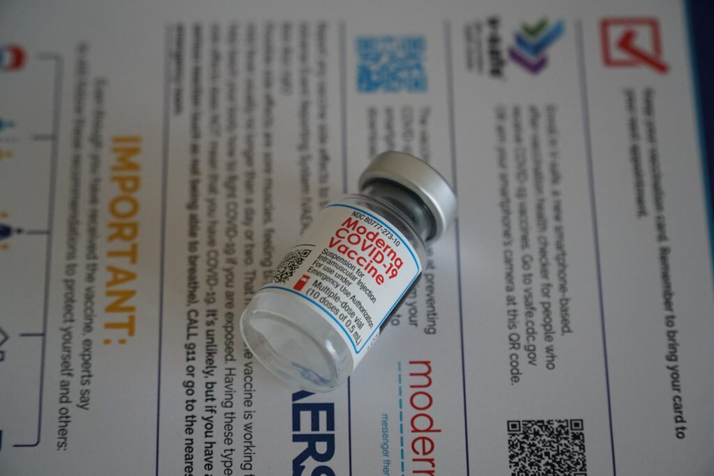
Toxicologist Janci Chunn Lindsay, Ph.D., calls on ACIP to halt Covid vaccine, citing fertility and blood clotting concerns. Photo of Moderna vaccine vial courtesy of Ian Hutchinson. | Jennifer Margulis, Ph.D.

In a public comment to the CDC, molecular biologist and toxicologist Dr. Janci Chunn Lindsay, Ph.D., called to immediately halt Covid vaccine production and distribution. Citing fertility, blood-clotting concerns (coagulopathy), and immune escape, Dr. Lindsay explained to the committee the scientific evidence showing that the coronavirus vaccines are not safe.

On April 23, 2021, the CDC’s Advisory Committee on Immunization Practices held a meeting in Atlanta, Georgia. The focus of this ACIP meeting was blood clotting disorders following Covid vaccines. Dr. Janci Chunn Lindsay spoke to the CDC during the time set aside for public comment.

The censorship on social media in particular and the internet in general is relentless. Here is a slightly edited, annotated censorship-proof transcript of Dr. Janci Chunn Lindsay’s 3-minute comment.

You can listen to her testimony on YouTube here (for now, anyway. If this link goes viral, YouTube will likely censor it).

Molecular Biologist and Toxicologist Calls to Halt Covid Vaccine

Hi, my name is Dr. Janci Chunn Lindsay. I hold a doctorate in biochemistry and molecular biology from the University of Texas, and have over 30 years of scientific experience, primarily in toxicology and mechanistic biology.

In the mid-1990s, I aided the development of a temporary human contraceptive vaccine which ended up causing unintended autoimmune ovarian destruction and sterility in animal test models. Despite efforts against this and sequence analyses that did not predict this.

I strongly feel that all the gene therapy vaccines must be halted immediately due to safety concerns on several fronts.

Janci Chunn Lindsay: Covid vaccines could induce cross-reactive antibodies to syncytin, and impair fertility as well as pregnancy outcomes

First, there is a credible reason to believe that the Covid vaccines will cross-react with the syncytin and reproductive proteins in sperm, ova, and placenta, leading to impaired fertility and impaired reproductive and gestational outcomes. 

Respected virologist Dr. Bill Gallaher, Ph.D., made excellent arguments as to why you would expect cross reaction. Due to beta sheet conformation similarities between spike proteins and syncytin-1 and syncytin-2.

I have yet to see a single immunological study which disproves this. Despite the fact that it would literally take the manufacturers a single day to do these syncytin studies to ascertain this [once they had serum from vaccinated individuals]. It’s been over a year since the assertions were first made that this [the body attacking its own syncytin proteins due to similarity in spike protein structure] could occur.

Pregnancy losses reported to VAERS lead to demand to halt Covid vaccine

We have seen 100 pregnancy losses reported in VAERS as of April 9th. And there have [also] been reports of impaired spermatogenesis and placental findings from both the natural infection, vaccinated, and syncytin knockout animal models that have similar placental pathology, implicating a syncytin-mediated role in these outcomes.

Additionally, we have heard of multiple reports of menses irregularities in those vaccinated. These must be investigated.

We simply cannot put these [vaccines] in our children who are at .002% risk for Covid mortality, if infected, or any more of the child-bearing age population without thoroughly investigating this matter.

[If we do], we could potentially sterilize an entire generation. Speculation that this will not occur and a few anecdotal reports of pregnancies within the trial are not sufficient proof that this is not impacting on a population-wide scale.

Covid vaccine causes blood disorders

Secondly, all of the gene therapies [Covid vaccines] are causing coagulopathy. [Coagulopathy when the body’s blood clotting system is impaired.] This is not isolated to one manufacturer. And this is not isolated to one age group. 

As we are seeing coagulopathy deaths in healthy young adults with no secondary comorbidities.

There have been 795 reports related to blood clotting disorders as of April 9th in the VAERS reporting system, 338 of these being due to thrombocytopenia.

There are forward and backward mechanistic principles for why this is happening. The natural infection is known to cause coagulopathy due to the spike protein. All gene therapy vaccines direct the body to make the spike protein. Zhang et al in [a scientific paper published in the Journal of Hematology & Oncology] in September 2020 showed that if you infuse spike protein into mice that have humanized ACE-2 receptors on blood platelets that you also get disseminated thrombosis.

Spike protein incubated with human blood in vitro also caused blood clot development which was resistant to fibrinolysis. [Fibrinolysis is the body’s process of breaking down blood clots]. The spike protein is causing thrombocytic events, which cannot be resolved through natural means. And all vaccines must be halted in the hope that they can be reformulated to guard against this adverse effect. 

Third, there is strong evidence for immune escape—

At this point in her oral testimony, Dr. Janci Chunn Lindsay was interrupted by a man’s voice: “Thank you for your comment, your time has expired.”

I reached out to Dr. Janci Chunn Lindsay to find out what else she had wanted to share with ACIP, in addition to her concerns over fertility and blood-clotting disorders. She sent me back her third point, which she submitted as written testimony.

Third, there is strong evidence for immune escape, and that inoculation under pandemic pressure with these leaky vaccines is driving the creation of more lethal mutants that are both newly infecting a younger age demographic, and causing more Covid-related deaths across the population than would have occurred without intervention. That is, there is evidence that the vaccines are making the pandemic worse.

It is clear that we are seeing a temporal immune depression immediately following the inoculations [see World Meter Global Covid deaths counts following inoculation dates] and there are immunosuppressive regions on spike proteins, as well as Syn-2, that could be likely causing this, through a T-cell mediated mechanism. If we do not stop this vaccine campaign until these issues can be investigated, we may see a phenomenon such as we see in chickens with Marek’s disease.

We have enough evidence now to see a clear correlation with increased Covid deaths and the vaccine campaigns. This is not a coincidence. It is an unfortunate unintended effect of the vaccines. We simply must not turn a blind eye and pretend this is not occurring. We must halt all Covid vaccine administration immediately, before we create a true pandemic that we cannot reign in.

MIT scientist also concerned about blood-clotting, fertility issues

Stephanie Seneff, Ph.D., an expert in protein synthesis, believes that Dr. Lindsay’s hypothesis is correct. “I absolutely share these concerns,” Dr. Seneff, who is a senior research scientist at MIT, wrote to me in a sobering email.

“The potential for blood clotting disorders and the potential for sterilization are only part of the story. There are other potential long-term effects of these vaccines as well, such as autoimmune disease and immune escape, whereby the vaccines administered to immune-compromised people accelerate the mutation rate of the virus so as to render both naturally acquired and vaccine-induced antibodies no longer effective.”

Like Dr. Lindsay, Dr. Seneff believes we need to immediately halt Covid vaccine campaigns. “This massive clinical trial on the general population could have devastating and irreversible effects on a huge number of people,” Seneff explains.

Despite these fertility and blood disorder concerns, the CDC panel voted last Friday to resume the use of the Johnson and Johnson vaccine. They did, however, suggest an FDA warning label be added. Their argument against halting Covid vaccination? The CDC believes the benefits outweigh the risks.

Dr. Janci Chunn Lindsay, Ph.D., calls on the CDC to halt Covid vaccine administration, citing fertility and blood-clotting concerns. | Jennifer Margulis, Ph.D.About Dr. Janci Chunn Lindsay

Dr. Janci Chunn Lindsay is the Director of Toxicology and Molecular Biology for Toxicology Support Services, LLC. She holds a doctorate in Biochemistry and Molecular Biology from the University of Texas Graduate School of Biomedical Sciences, M.D. Anderson Cancer Center-Houston. Dr. Lindsay has extensive experience in analyzing the molecular profile of pharmacologic responses. Her expertise centers on evaluating the complex dynamics of toxicity, such as toxicant pharmacology, exposure route, host metabolism, and subsequent cellular effects as they relate to the contribution of specific substances to impairment, health and fertility risk, and human disease.

Source:https://rightsfreedoms.wordpress.com/2021/05/06/halt-covid-vaccine-prominent-scientist-tells-cdc/

Παρά τις εφιαλτικές προειδοποιήσεις των ειδικών η πολιτικο-οικονομικο-υγειονομική χούντα συνεχίζει αδίστακτη τους φόνους αθώων ανενημέρωτων πολιτών

Δρ. Janci Chunn Lindsay: Τὰ ἐμβόλια προκαλοῦν στειρώσεις, θρομβώσεις καὶ περισσότερες θανατηφόρες μεταλλάξεις τοῦ ἰοῦ!

Ἡ Δρ. Janci Chunn Lindsay εἶναι διακεκριμένη ἐπιστήμονας στὸν τομέα τῆς τοξικολογίας. Εἶναι διδάκτωρ τῆς Βιοχημείας καὶ τῆς Μοριακῆς Βιολογίας τοῦ Πανεπιστημίου τοῦ Τέξας, Η.Π.Α. καὶ γιὰ περισσότερα ἀπὸ 30 χρόνια διεξάγει ἐπιστημονικές ἔρευνες στὸν τομέα τῆς τοξικογονιδιωματικῆς. Ἡ τεχνογνωσία της ἐπικεντρώνεται στὶς βλάβες ποὺ προκαλοῦν διάφορες οὐσίες στὴν ἀνθρώπινη ὑγεία. Ἐπιπλέον διενεργεῖ ἰατροδικαστικὲς τοξικολογικὲς ἔρευνες.

Οἱ βασικὲς θέσεις τῆς Δρ. Lindsay ἀναφορικὰ μὲ τὰ ἐμβόλια Covid-19:

  • Τὰ ἐμβόλια Covid-19 εἶναι πολὺ πιθανὸ νὰ ἀντιδράσουν μὲ τὴ συγκυτίνη καθὼς ἐπίσης μὲ ἄλλες ἀναπαραγωγικὲς πρωτεΐνες στὸ σπέρμα, τὰ ὠάρια καὶ τὸν πλακούντα, ὁδηγώντας ἔτσι σὲ μειωμένη γονιμότητα καὶ μειωμένα ἀποτελέσματα ἀναπαραγωγῆς καὶ κύησης.
  • Δὲν ἐνδείκνυται νὰ χορηγήσουμε αὐτὰ τὰ ἐμβόλια στὰ παιδιὰ ἤ σὲ ὁποιονδήποτε ἄλλο πληθυσμὸ ἀναπαραγωγικῆς ἡλικίας. Ἐὰν τὸ κάνουμε, ἐνδεχομένως νὰ στειρώσουμε μία ὁλόκληρη γενιά.
  • Ὅλα τὰ γενετικὰ ἐμβόλια Covid-19 προκαλοῦν θρομβώσεις. Αὐτὸ δὲν ἀφορᾶ μόνο μία ἡλικιακὴ ὁμάδα. Βλέπουμε θανάτους λόγῳ θρόμβωσης σὲ ἐμβολιασμένους ὑγιεῖς νεαροὺς ἐνήλικες.
  • Ὁ ἐμβολιασμὸς κατὰ τὴ διάρκεια πανδημίας μὲ τὰ συγκεκριμένα ἐμβόλια ὁδηγεῖ στὴ δημιουργία περισσότερων θανατηφόρων μεταλλάξεων, πού μολύνουν νεαρότερες ἡλικίες καὶ προκαλοῦν περισσότερους θανάτους ἀπὸ ὅσους θὰ εἴχαμε, ἐὰν δὲν εἶχαν γίνει οἱ ἐμβολιασμοί. Τὰ ἐμβόλια ἐπιδεινώνουν τὴν πανδημία.


Ἡ Δρ. Lindsay στέλνει μία δραματικὴ ἔκκληση: «Πρέπει νὰ σταματήσουμε ἀμέσως κάθε χορήγηση ἐμβολίων Covid-19, προτοῦ δημιουργήσουμε μία πραγματικὴ πανδημία τὴν ὁποία δὲν θὰ μποροῦμε νὰ ἐλέγξουμε».

Πηγὲς

  • https://e-deltio.com/data/files/2.pdf
  • https://rightsfreedoms.wordpress.com/2021/05/06/halt-covid-vaccine-prominent-scientist-tells-cdc/

Γονείς, σταματείστε να δολοφονείτε τα παιδιά σας υπακούοντας στα ψέματα που σας προβάλουν τα άθλια κανάλια και η world mafia! Μασονία, Σιωνισμός Ιλλουμινάτοι, Ροδόσταυροι, Sculls and Βones και η Λέσχη Μπίλντεμπεργκ

Δραματικὴ αὔξηση τῆς παιδικῆς θνησιμότητας στὴ Βρετανία μετὰ ἀπὸ τοὺς ἐμβολιασμοὺς

Στὶς 13 Σεπτεμβρίου 2021 ὁ Chris Whitty, ὁ Ἐπικεφαλῆς Ἰατρικὸς Λειτουργὸς τῆς Ἀγγλίας, συμβούλευσε τὴν κυβέρνηση τῆς Βρετανίας νὰ ἐμβολιάσει ὅλα τὰ παιδιὰ ἡλικίας ἄνω τῶν 12 ἐτῶν μὲ τουλάχιστον μία δόση τοῦ ἐμβολίου Pfizer Covid-19. Ἡ Κοινὴ Ἐπιτροπὴ Ἐμβολιασμῶν καὶ Ἀνοσοποίησης (JCVI) εἶχε ἀντίθετη ἄποψη, διότι εἶναι ἀμελητέος ὁ ἀριθμὸς τῶν παιδιῶν ποὺ ὑποφέρουν ἀπὸ σοβαρὴ ἀσθένεια λόγῳ Covid-19, ἐνῶ εἶναι ὑπαρκτὴ ἡ πιθανότητα τὰ παιδιὰ νὰ ὑποστοῦν μυοκαρδίτιδα λόγῳ τοῦ ἐμβολίου. Ἡ βρετανικὴ κυβέρνηση ἀγνόησε τὶς συμβουλὲς τοῦ JCVI καὶ ξεκίνησε ἀμέσως τὸν ἐμβολιασμὸ τῶν παιδιῶν ἄνω τῶν 12 ἐτῶν. Τὸ ἀποτέλεσμα: Αὐξήθηκε ἡ γενικὴ θνησιμότητα τῶν παιδιῶν, δηλαδὴ οἱ θάνατοι ἀπὸ κάθε αἰτία. Τὰ στοιχεῖα τοῦ πιὸ κάτω πίνακα λήφθηκαν ἀπὸ τὴν Ἐθνικὴ Στατιστικὴ Ὑπηρεσία (ONS – Office of National Statistics) τῆς Βρετανίας.

Θάνατοι παιδιῶν 10–14 ἐτῶν στὴν Ἀγγλία καὶ Οὐαλλία
ΕΤΟΣ 2015–2019 (μέσος ὅρος)1 20202 20213
Θάνατοι ἀπὸ κάθε αἰτία ἐκτὸς κορωνοϊοῦ 41 43 55
Θάνατοι σχετιζόμενοι μὲ κορωνοϊὸ4 0 0 4
Σύνολο 41 43 59

1 38η μέχρι 45η ἑβδομάδα (δὲν δίνονται ἡμερομηνίες ἀπὸ τὸ ONS)

2 39η μέχρι 46η ἑβδομάδα (25 Σεπτεμβρίου – 19 Νοεμβρίου 2020)

3 38η μέχρι 45η ἑβδομάδα (24 Σεπτεμβρίου – 18 Νοεμβρίου 2021)

4 Σχετιζόμενοι (involving Covid-19), ὄχι λόγῳ κορωνοϊού (due to Covid-19)

Τὸ 2021 (μετὰ τὴν ἔναρξη τῶν μαζικῶν ἐμβολιασμῶν τῶν παιδιῶν) καταγράφηκαν 12 θάνατοι (ἀπὸ κάθε αἰτία ἐκτὸς κορωνοϊοῦ) περισσότεροι ἀπὸ τὸ 2020 (αὔξηση 27.9%) καὶ 14 θάνατοι (ἀπὸ κάθε αἰτία ἐκτὸς κορωνοϊοῦ) περισσότεροι ἀπὸ τὸν μέσο ὅρο τῶν ἐτῶν 2015–2019 (αὔξηση 34.1%). Ἡ μόνη γνωστὴ καὶ μετρήσιμη ὑγειονομικὴ ἀλλαγὴ ποὺ ἐπῆλθε στὴ ζωὴ τῶν παιδιῶν τῆς Βρετανίας ἀπὸ τὴν 38η ἑβδομάδα τοῦ 2021 καὶ μετὰ εἶναι ὁ μαζικὸς ἐμβολιασμὸς μὲ τὸ ἐμβόλιο Pfizer Covid-19. Εἶναι λογικὸ νὰ θεωροῦμε ὅτι ὑπάρχουν πολλὲς πιθανότητες αὐτὸς ὁ ἐμβολιασμὸς νὰ εὐθύνεται γιὰ τὴ δραματικὴ αὔξηση τῆς γενικῆς παιδικῆς θνησιμότητας στὴ Βρετανία.

Πηγὴ

https://dailyexpose.uk/2021/11/24/child-deaths-increasing-since-offered-covid-vaccine/

Μπράβο βραβευμένε Μπουρλά σκότωσε κι άλλους με την εταιρεία σου. Πήρες επάξιο συγχωροχάρτι από τους μασονοσιωνιστές!

Το CNN βράβευσε τον Albert Bourla της Pfizer ως CEO της Χρονιάς. Η Νότια Κορέα παρουσιάζει επίσημα την αιτιότητα του εμβολιασμού κατά της COVID-19 και της μυοκαρδίτιδας

Tα Κέντρα Ελέγχου και Πρόληψης Νοσημάτων της Νοτίου Κορέας αναγνώρισαν την αιτιότητα του εμβολιασμού κατά του COVID-19 και της μυοκαρδίτιδας

Το CNN βράβευσε τον CEO της Pfizer ως τον CEO της Χρονιάς. Eκείνος, ο Albert Bourla, ανταποκρίθηκε ως εξής>

Σ.γ.:NOBEL δεν θα του απονείμετε; Αυτός και οι συμμορίτες συνάδελφοί του ΣΩΖΟΥΝ ΛΑΟΥΣ

«Τι αξιοσημείωτη τιμή από το CNN καθώς πλησιάζουμε στο τέλος μιας πολυάσχολης, απαιτητικής και επιτυχημένης χρονιάς για την Pfizer. Μια χρονιά που δεν θα ξεχάσω ποτέ. Ευχαριστώ όλους τους συναδέλφους μας. Είναι ο λόγος που έχω απολαύσει κάθε λεπτό των 28 χρόνων μου εδώ».

Ανάμεσα στους διεκδικητές του τίτλου υπήρξαν η Lisa Su της AMD, ο Jim Farley της Ford, ο Marvin Ellison της Lowe’s, ο David Solomon της Goldman Sachs, ο Sundar Pichai της Alphabet και βέβαια, ο Elon Musk των Tesla και SpaceX, ο οποίος έλαβε τον τίτλο της προσωπικότητας της χρονιάς τόσο από το περιοδικό Time όσο και από τους Financial Times.

«Όμως ο Μπουρλά ήταν η δική μας επιλογή, για όσα έχει κάνει η Pfizer σε έναν χρόνο όπου τα εμβόλια κατά του κορωνοϊού έγιναν mainstream (το ‘εμβόλιο’ ξεχώρισε ως η λέξη της χρονιάς από το Merriam-Webster) και βοήθησαν να σταθεροποιηθεί η αμερικανική οικονομία – και στην πραγματικότητα, η παγκόσμια», αναφέρει το CNN.

«Η Pfizer έκανε πολύ καλό για την ανθρωπότητα και είμαστε πολύ, πολύ υπερήφανοι για αυτό», είπε ο Bourla σε συνέντευξή του στο CNN Business. «Δεν μπορέσαμε μόνο να σώσουμε τόσο πολλές ζωές. Αλλά απολαμβάνουμε υψηλά επίπεδα επιχειρηματικής φήμης αυτή τη στιγμή. Οι άνθρωποι μας συμπαθούν».

Όπως εξηγεί το CNN, η Pfizer δεν είναι η μοναδική εταιρεία που ανέπτυξε εμβόλιο κατά του κορωνοϊού. Όμως αυτός ο ηγέτης του Big Pharma έχει γίνει συνώνυμο της μάχης απέναντι στον κορωνοϊό, περισσότερο από την BioNTech με την οποία συνεργάστηκε ή από τη Moderna.

Σε ό,τι αφορά την εξάπλωση της νέας μετάλλαξης, ο Βourla είπε στο CNN ότι το εμβόλιο των  Pfizer/ BioNTech θα ελέγξει την Όμικρον «αρκετά καλά», ειδικά εάν ο κόσμος λάβει τρεις δόσεις.

Όμως παραδέχθηκε ότι ενδεχομένως η εταιρεία να χρειαστεί να κυκλοφορήσει ένα νέο εμβόλιο, που να στοχεύει ειδικά στην Όμικρον ή σε τυχόν μελλοντικά στελέχη. Ήδη η εταιρεία αναπτύσσει ένα τέτοιο εμβόλιο, το οποίο ο ίδιος εκτιμά ότι θα τεθεί προς έγκριση έως τον Μάρτιο.

Tα Κέντρα Ελέγχου και Πρόληψης Νοσημάτων της Νοτίου Κορέας αναγνώρισαν την αιτιότητα του εμβολιασμού κατά του COVID-19 και της μυοκαρδίτιδας.  

Ο αποθανών ήταν 22χρονος νεοσύλλεκτος στρατιωτικός που έφυγε από τη ζωή 6 μέρες μετά τον εμβολιασμό του με το εμβόλιοmRNA BNT162b2 της pfizer.

Η ΑΝΑΤΟΜΙΑ, ΤΑ ΕΥΡΗΜΑΤΑ, ΤΟ ΣΥΜΠΕΡΑΣΜΑ

Παρουσιάζουμε τώρα τα ευρήματα αυτοψίας ενός 22χρονου άνδρα που εμφάνισε πόνο στο στήθος 5 ημέρες μετά την πρώτη δόση του εμβολίου mRNA BNT162b2 και πέθανε 7 ώρες αργότερα.

Αυτή είναι η πρώτη περίπτωση στη Νότια Κορέα που τα Κέντρα Ελέγχου και Πρόληψης Νοσημάτων της Κορέας αναγνώρισαν την αιτιότητα του εμβολιασμού κατά του COVID-19 και της μυοκαρδίτιδας. Αυτή η μοναδική περίπτωση αποτελεί παράδειγμα σοβαρού ανεπιθύμητου συμβάντος μετά τον εμβολιασμό με mRNA κατά του COVID-19.

Η ιστολογική εξέταση της καρδιάς αποκάλυψε μεμονωμένη κολπική μυοκαρδίτιδα, με υπεροχή ουδετερόφιλων και ιστιοκυττάρων. Η ανοσοϊστοχημική χρώση C4d αποκάλυψε διάσπαρτη μονοκυτταρική νέκρωση των μυοκυττάρων η οποία δεν συνοδεύτηκε από φλεγμονώδεις διηθήσεις. Παρατηρήθηκε εκτεταμένη νέκρωση ζώνης συστολής στους κόλπους και τις κοιλίες. Δεν υπήρχαν ενδείξεις μικροθρόμβωσης ή μόλυνσης στην καρδιά και σε άλλα όργανα. Η κύρια αιτία θανάτου προσδιορίστηκε ότι ήταν η μυοκαρδίτιδα, που σχετίζεται αιτιολογικά με το εμβόλιο BNT162b2.

Η πανδημία της νόσου του κορωνοϊού 2019 (COVID-19), που ξεκίνησε τον Ιανουάριο του 2020, έχει επηρεάσει ανθρώπους σε όλο τον κόσμο. Η ταχεία ανάπτυξη εμβολίου επέτρεψε την πρόληψη του COVID-19. Δύο εμβόλια mRNA COVID-19, BNT162b2 (Pfizer-BioNTech) και mRNA-1273 (Moderna) επέδειξαν ασφάλεια και αποτελεσματικότητα σε κλινικές δοκιμές και τους χορηγήθηκε άδεια χρήσης έκτακτης ανάγκης από την Υπηρεσία Τροφίμων και Φαρμάκων των ΗΠΑ τον Δεκέμβριο του 2020. Τα άτομα που εμβολιάστηκαν κατά του COVID-19 έχει αυξηθεί, έχουν υπάρξει πολλαπλές αναφορές μυοκαρδίτιδας ως ανεπιθύμητο συμβάν μετά από ανοσοποίηση, που δεν παρατηρήθηκε σε κλινικές δοκιμές.

Η μυοκαρδίτιδα χαρακτηρίζεται ιστολογικά από διάχυτες και/ή εστιακές φλεγμονώδεις διηθήσεις εντός του μυοκαρδιακού ιστού, συνοδευόμενες από βλάβη των μυοκυττάρων χωρίς ενδείξεις ισχαιμίας.

Λίγα είναι γνωστά για τα ιστοπαθολογικά χαρακτηριστικά της μυοκαρδίτιδας μετά τον εμβολιασμό για τον COVID-19, επειδή η βιοψία του ενδομυοκαρδίου δεν είναι μια διαδικασία ρουτίνας για τη μυοκαρδίτιδα και, λόγω της γενικά ευνοϊκής πρόγνωσής του, ευκαιρίες για αυτοψικές μελέτες είναι σπάνιες.

Παρατηρήσαμε πρόσφατα μια περίπτωση αιφνίδιου καρδιακού θανάτου σε νεαρό άνδρα έξι ημέρες μετά τη λήψη της πρώτης δόσης του εμβολίου mRNA BNT162b2 και αναφέραμε τα διακριτικά κλινικά και παθολογικά ευρήματα αυτής της περίπτωσης.

Ο αποθανών ήταν 22χρονος νεοσύλλεκτος στρατιωτικός.

Η αρτηριακή του πίεση ήταν αυξημένη κατά τη φυσική εξέταση 17 και 7 μήνες πριν από το θάνατό του (156/94 mmHg και 128/74 mmHg, αντίστοιχα), αλλά κατά τα άλλα ήταν υγιής.

Στις 13 Ιουνίου 2021, 5 ημέρες μετά την πρώτη δόση του εμβολιασμού mRNA BNT162b2, παραπονέθηκε σε έναν συνάδελφό του για πόνο στο στήθος στη 1:00 π.μ., κατά τη διάρκεια ενός διαλείμματος  και πήγε για ύπνο. Στις 8:00 το πρωί βρέθηκε αναίσθητος καμπουριασμένος δίπλα στο κρεβάτι. Μεταφέρθηκε σε τμήμα επειγόντων περιστατικών και στο ηλεκτροκαρδιογράφημα διαπιστώθηκε κοιλιακή μαρμαρυγή. Έγινε καρδιοπνευμονική ανάνηψη για δύο ώρες, αλλά δεν μπόρεσε να αναζωογονηθεί.

Διενεργήθηκε νεκροψία-νεκροτομή 24 ώρες μετά τον θάνατό του. Ο θανών ήταν καλά σιτισμένος χωρίς εμφανή τραύματα στην εξωτερική εξέταση. Η καρδιά ζύγιζε 470 g και είχε πολλαπλές πετέχειες στην επιφάνειά της. Το περικάρδιο ήταν λείο χωρίς εναπόθεση ινώδους ή εξίδρωμα. Οι στεφανιαίες αρτηρίες ήταν λανθάνουσες και οι καρδιακές βαλβίδες δεν ήταν αξιοσημείωτες. Το μυοκάρδιο ήταν φυσιολογικού πάχους και δεν υπήρχε διαστολή των κόλπων ή των κοιλιών. Το μυοκάρδιο ήταν ομοιογενώς καφέ χωρίς εμφανή νέκρωση ή ίνωση.

Στη μικροσκοπική εξέταση, διάχυτη φλεγμονώδης διήθηση, με κυριαρχία ουδετερόφιλων και ιστιοκυττάρων, παρατηρήθηκε εντός του μυοκαρδίου. Συγκεκριμένα, τα φλεγμονώδη διηθήματα ήταν κυρίαρχα στους κόλπους  και γύρω από τους φλεβοκομβικούς (SA) και κολποκοιλιακούς (AV) κόμβους  ενώ η κοιλιακή περιοχή εμφάνιζε ελάχιστα ή καθόλου φλεγμονώδη κύτταρα.

Περιστασιακή νέκρωση ή εκφυλισμός μυοκυττάρων βρέθηκε δίπλα στα φλεγμονώδη διηθήματα, χωρίς σχηματισμό αποστήματος ή βακτηριακό αποικισμό. Υπήρχε επίσης διάσπαρτη μονοκυτταρική νέκρωση μυοκυττάρων χωρίς συνοδό φλεγμονή. Πολλαπλές διάσπαρτες εστίες νέκρωσης ζώνης συστολής (CBN) αναγνωρίστηκαν σε όλο το μυοκάρδιο, κυρίως στην αριστερή κοιλία. Δεν βρέθηκαν άλλες συγκεκριμένες παθολογικές αλλαγές στον πνεύμονα, το ήπαρ, τα νεφρά, τον σπλήνα, το πάγκρεας ή τον εγκέφαλο σε μακροσκοπική ή μικροσκοπική εξέταση.
Αιτία θανάτου προσδιορίστηκε η μυοκαρδίτιδα. Δεδομένου ότι η μυοκαρδίτιδα έδειξε μια χρονική σχέση με τη χορήγηση του εμβολίου και δεν υπήρχε άλλη εξήγηση για τον αιφνίδιο καρδιακό θάνατο, στις 26 Ιουλίου 2021, τα Κέντρα Ελέγχου και Πρόληψης Νοσημάτων της Κορέας αναγνώρισαν ότι η μυοκαρδίτιδα και ο εμβολιασμός «πιθανώς σχετίζονται» σε αυτήν την περίπτωση.

Δήλωση ηθικής

Λήφθηκε ενημερωμένη συγκατάθεση για δημοσίευση κλινικών δεδομένων από την οικογένεια του θανόντος.

>Υπήρχαν τρία κύρια ιστολογικά ευρήματα στην καρδιά: 1) μυοκαρδίτιδα που αφορούσε κυρίως το τοίχωμα των κόλπων, με υπεροχή ουδετερόφιλων και ιστιοκυττάρων.

2) μη φλεγμονώδης μονοκυτταρική νέκρωση.

και 3) διάχυση CBN σε όλο το μυοκάρδιο, κυρίως στην αριστερή κοιλία. Αυτά τα παθολογικά ευρήματα δεν ήταν εμφανή στη μακροσκοπική εξέταση. Το μόνο μη φυσιολογικό χονδρό εύρημα ήταν η καρδιακή διεύρυνση, η οποία μπορεί να ήταν δευτερογενής στην υπέρταση.

Σε αυτή την περίπτωση, η μυοκαρδίτιδα ήταν ιστολογικά διαφορετική από την ιογενή ή ανοσο-μεσολαβούμενη μυοκαρδίτιδα στο ότι τα φλεγμονώδη διηθήματα ήταν κυρίως ουδετερόφιλα και ιστιοκύτταρα, παρά λεμφοκύτταρα. Δεν παρατηρήθηκαν πολυπύρηνα γιγαντιαία κύτταρα. Η περιοχή της μυοκαρδίτιδας περιοριζόταν στο κολπικό τοίχωμα, αλλά οι κοιλίες ήταν απαλλαγμένες από κυτταρική διήθηση. Φλεγμονώδη κύτταρα υπήρχαν επίσης στους κόμβους SA και AV. Παρατηρήθηκε νέκρωση ή εκφυλισμός μυοκυττάρων δίπλα στα φλεγμονώδη διηθήματα. Η διήθηση ουδετερόφιλων του μυοκαρδίου είναι ένας ασυνήθιστος ιστολογικός τύπος μυοκαρδίτιδας και γενικά παρατηρείται σε ανοσοκατεσταλμένους ασθενείς με βακτηριακή λοίμωξη.

Ο εκλιπών ήταν προηγουμένως υγιής και δεν υπήρχαν σημεία λοιμώδους μυοκαρδίτιδας. Αποκλείστηκε επίσης η διάδοση λοιμώδους ενδοκαρδίτιδας και πνευμονίας. Ο υποκείμενος μηχανισμός της βλάβης του μυοκαρδίου σε αυτή την περίπτωση είναι ασαφής, αλλά μπορεί να εμπλέκεται ανοσολογική βλάβη του μυοκαρδίου που προκαλείται από κυτοκίνη ή συνδέεται με ιστιοκύτταρα. Η μεμονωμένη κολπική μυοκαρδίτιδα είναι πολύ σπάνια και επομένως είναι μια άγνωστη οντότητα ασθένειας στους παθολόγους. Από ό,τι γνωρίζουμε, μόνο δύο αναφορές περιστατικών αιφνίδιου καρδιακού θανάτου που προκαλείται από μεμονωμένη κολπική μυοκαρδίτιδα έχουν δημοσιευτεί στο παρελθόν.

Προηγούμενες αναφορές σημείωσαν ότι η κολπική μυοκαρδίτιδα μπορεί να αγνοηθεί κατά τη μεταθανάτια εξέταση, επειδή η δειγματοληψία κόλπου δεν πραγματοποιείται τακτικά. Στην παρούσα περίπτωση, η εκτεταμένη δειγματοληψία μυοκαρδίου επέτρεψε την ακριβή διάγνωση. Συνολικά εξετάστηκαν 35 τομές (25 τμήματα κοιλιακού και κολπικού μυοκαρδίου και 10 τμήματα του συστήματος αγωγής) και 9 τομές είχαν ενδείξεις μυοκαρδίτιδας. Η μυοκαρδίτιδα συνήθως δεν είναι εμφανής κατά τη συνολική εξέταση. Ακόμα κι αν η καρδιά είναι κατάφωρα μη αξιοσημείωτη, οι παθολόγοι θα πρέπει να εξετάσουν επαρκή αριθμό (≥ 10) κολπικών και κοιλιακών τομών για να μην παραβλέψουν ή να διαγνώσουν εσφαλμένα την αιτία θανάτου, ειδικά εάν ο θανών είχε συμπτώματα μυοκαρδίτιδας και πρόσφατο ιστορικό εμβολιασμού mRNA.

Σημειωτέον, μονοκυτταρική νέκρωση (ή μονοκυτταρική ισχαιμία) των μυοκυττάρων χωρίς φλεγμονή παρατηρήθηκε σε πολλαπλές θέσεις σε όλους τους κόλπους. Αρκετές μελέτες αυτοψίας των καρδιαγγειακών παθολογικών ευρημάτων ατόμων που πέθαναν από COVID-19 έχουν επίσης βρει μονοκυτταρική νέκρωση και αυτό μπορεί να είναι ένα διακριτικό παθολογικό χαρακτηριστικό του COVID-19. Έτσι, τραυματισμός του μυοκαρδίου λόγω εμβολιασμού κατά του COVID-19 μπορεί ιστολογικά να παρουσιαστεί όχι μόνο ως μυοκαρδίτιδα, αλλά και ως διάσπαρτη μονοκυτταρική νέκρωση, παρόμοια με τις μυοκαρδιακές βλάβες του COVID-19

Ένα άλλο σημαντικό ιστολογικό εύρημα ήταν το άφθονο CBN σε όλο το μυοκάρδιο, ειδικά στην αριστερή κοιλία. Η CBN συνήθως παρατηρείται μετά από μη αναστρέψιμο τραυματισμό των μυοκυττάρων και σχετίζεται με ισχαιμική καρδιακή νόσο ή κατάσταση περίσσειας κατεχολαμινών.

Η CBN σε αυτή την περίπτωση ήταν εκτεταμένη σε όλο το κοιλιακό μυοκάρδιο και είμαστε προσεκτικοί ως προς την εξαγωγή συμπερασμάτων σχετικά με την αιτιότητα. Η συσχέτιση μεταξύ του εμβολιασμού CBN και του COVID-19 είναι ασαφής. Το CBN μπορεί να έχει συμβεί ως αποτέλεσμα κοιλιακής μαρμαρυγής ή χορήγησης κατεχολαμίνης κατά τη διάρκεια της ανάνηψης. Δεν υπήρχαν ενδείξεις αθηροσκλήρωσης ή μικροθρόμβωσης των στεφανιαίων, που θα μπορούσαν να εξηγήσουν την παρουσία CBN.

Μπορέσαμε να βρούμε μόνο μία προηγουμένως δημοσιευμένη αναφορά περιστατικού θανάτου λόγω μυοκαρδίτιδας μετά από εμβολιασμό κατά του COVID-19 με μια ολοκληρωμένη κλινικοπαθολογική ανάλυση. Οι Verma et al.10 ανέφεραν την περίπτωση ενός άνδρα 42 ετών, ο οποίος παρουσίασε πόνο στο στήθος και δύσπνοια 2 εβδομάδες μετά τη δεύτερη δόση του εμβολιασμού mRNA-1273 και πέθανε 3 ημέρες μετά την έναρξη των συμπτωμάτων. Στη μικροσκοπική εξέταση, η βλάβη των μυοκυττάρων με μικτό φλεγμονώδες διήθημα μακροφάγων, λεμφοκυττάρων και ηωσινόφιλων παρατηρήθηκε στο μυοκάρδιο και των δύο κοιλιών, ενώ οι κόλποι δεν έδειξαν στοιχεία μυοκαρδίτιδας.

Αντίθετα, ο ασθενής της περίπτωσής μας εμφάνισε συμπτώματα 5 ημέρες μετά την πρώτη δόση του εμβολίου mRNA BNT162b2 και πέθανε 7 ώρες αργότερα και εμφάνισε μεμονωμένη κολπική μυοκαρδίτιδα με υπεροχή ουδετερόφιλων και ιστιοκυττάρων. Τα ιστοπαθολογικά χαρακτηριστικά που περιγράφονται στην αναφορά των Verma et al.10 δεν περιελάμβαναν CBN ή μονοκυτταρική νέκρωση των μυοκυττάρων. Αυτό υποδηλώνει ότι η μυοκαρδίτιδα μετά τον εμβολιασμό με mRNA του COVID-19 είναι ετερογενής, τόσο κλινικά όσο και ιστολογικά.

Η μυοκαρδίτιδα που σχετίζεται με το εμβόλιο έχει αναφερθεί κυρίως σε νεαρούς άνδρες μετά τον δεύτερο εμβολιασμό. Η μυοκαρδίτιδα διαγνώστηκε κυρίως κλινικά με βάση την αυξημένη τροπονίνη ορού και την απεικόνιση καρδιακού μαγνητικού συντονισμού και όλοι οι ασθενείς εκτός από τον δικό μας και τον ασθενή που αναφέρθηκε από τους Verma et al.10 ανάρρωσαν μετά από υποστηρικτική φροντίδα.

Ο ασθενής σε αυτή την περίπτωση εμφάνισε παρόμοια κλινικά χαρακτηριστικά, γεγονός που υποστηρίζει μια πιθανή συσχέτιση μεταξύ εμβολιασμού και μυοκαρδίτιδας.

Ωστόσο, δεδομένου ότι άλλες αναφερόμενες περιπτώσεις μυοκαρδίτιδας ήταν γενικά ήπιες και τα συμπτώματα συνήθως εμφανίζονταν μετά τον δεύτερο εμβολιασμό, η κλινική πορεία της περίπτωσής μας (αιφνίδιος θάνατος 6 ημέρες μετά τον πρώτο εμβολιασμό) είναι ένα εξαιρετικά σπάνιο συμβάν. Λαμβάνοντας υπόψη το μικρό χρονικό διάστημα μεταξύ της έναρξης των συμπτωμάτων του αποθανόντος ασθενούς και του αιφνίδιου θανάτου του, η άμεση αιτία θανάτου ήταν πιθανώς μια αρρυθμία και όχι η καρδιακή ανεπάρκεια.

Αυτή είναι η πρώτη περίπτωση στη Νότια Κορέα που τα Κέντρα Ελέγχου και Πρόληψης Νοσημάτων της Κορέας αναγνώρισαν την αιτιότητα του εμβολιασμού κατά του COVID-19 και της μυοκαρδίτιδας. Αυτή η μοναδική περίπτωση αποτελεί παράδειγμα σοβαρού ανεπιθύμητου συμβάντος μετά τον εμβολιασμό με mRNA κατά του COVID-19. Δεν είναι γνωστό εάν αυτό το κρούσμα σχετίζεται με τον τύπο του εμβολίου ή με ένα συγκεκριμένο συστατικό του εμβολίου. Δεν είναι επίσης σαφές εάν η εντόπιση (κόλπος), ο τύπος της φλεγμονής (ουδετερόφιλα και ιστιοκύτταρα), η CBN και η μονοκυτταρική νέκρωση χωρίς φλεγμονή είναι ειδικά χαρακτηριστικά της μυοκαρδίτιδας που σχετίζεται με το εμβόλιο COVID-19. Απαιτείται ολοκληρωμένη κλινική και παθολογική αξιολόγηση πρόσθετων περιπτώσεων για να αποσαφηνιστεί η σχέση μεταξύ του εμβολιασμού κατά του COVID-19 και της μυοκαρδίτιδας.

ΠΗΓΗ:https://kourdistoportocali.com/news-desk/to-cnn-vraveyse-ton-albert-bourla-tis-pfizer-os-ceo-tis-chronias-i-notia-korea-paroysiazei-episima-tin-aitiotita-toy-emvoliasmoy-kata-tis-covid-19-kai-tis-myokarditidas/

 
2021 Oct 18; 36(40): e286.
Published online 2021 Jul 10. doi: 10.3346/jkms.2021.36.e286
PMCID: PMC8524235
PMID: 34664804

Myocarditis-induced Sudden Death after BNT162b2 mRNA COVID-19 Vaccination in Korea: Case Report Focusing on Histopathological Findings

Abstract

We present autopsy findings of a 22-year-old man who developed chest pain 5 days after the first dose of the BNT162b2 mRNA vaccine and died 7 hours later. Histological examination of the heart revealed isolated atrial myocarditis, with neutrophil and histiocyte predominance. Immunohistochemical C4d staining revealed scattered single-cell necrosis of myocytes which was not accompanied by inflammatory infiltrates. Extensive contraction band necrosis was observed in the atria and ventricles. There was no evidence of microthrombosis or infection in the heart and other organs. The primary cause of death was determined to be myocarditis, causally-associated with the BNT162b2 vaccine.

Keywords: Sudden Cardiac Death, Myocarditis, Coronavirus Disease 19, Vaccination, mRNA Vaccines, Adverse Event Following Immunization

Graphical Abstract

An external file that holds a picture, illustration, etc. Object name is jkms-36-e286-abf001.jpg


INTRODUCTION

The coronavirus disease 2019 (COVID-19) pandemic, which started in January 2020 has affected people worldwide. Rapid vaccine development has enabled COVID-19 prevention. Two mRNA COVID-19 vaccines, BNT162b2 (Pfizer-BioNTech) and mRNA-1273 (Moderna) demonstrated safety and efficacy in clinical trials, and were granted emergency use authorization by the US Food and Drug Administration in December 2020., As the number of people vaccinated against COVID-19 has increased, there have been multiple reports of myocarditis as an adverse event following immunization,,,,,,,,,, which was not observed in clinical trials.

Myocarditis is histologically characterized by diffuse and/or focal inflammatory infiltrates within myocardial tissue, accompanied by myocyte damage without evidence of ischemia. Little is known about the histopathological characteristics of myocarditis following COVID-19 vaccination because endomyocardial biopsy is not a routine procedure for myocarditis and, due to its generally favorable prognosis, opportunities for autopsy studies are rare.

We recently observed a case of sudden cardiac death in a young male six days after receiving the first dose of the BNT162b2 mRNA vaccine and report the distinctive clinical and pathological findings of this case.

CASE DESCRIPTION

The deceased was 22-year-old male military recruit. His blood pressure was elevated on physical examination 17 and 7 months before his death (156/94 mmHg and 128/74 mmHg, respectively), but he was otherwise healthy. On June 13, 2021, 5 days after the first dose of BNT162b2 mRNA vaccination, he complained to a colleague of chest pain at 1:00 AM, during a smoke break, and went to bed. At 8:00 AM, he was found unconscious hunched beside the bed. He was taken to an emergency department and was found to have ventricular fibrillation on electrocardiography. Cardiopulmonary resuscitation was performed for two hours, but he could not be resuscitated.

An autopsy was performed 24 hours after his death. The deceased was well-nourished with no visible injuries on external examination. The heart weighed 470 g and had multiple petechiae on its surface. The pericardium was smooth with no fibrin deposition or exudate. The coronary arteries were patent, and the heart valves were unremarkable. The myocardium was of normal thickness and there was no dilation of the atria or ventricles. The myocardium was homogeneously brown with no obvious necrosis or fibrosis. On microscopic examination, diffuse inflammatory infiltration, with neutrophil and histiocyte predominance, was observed within the myocardium (Fig. 1A). Notably, the inflammatory infiltrates were dominant in the atria (Fig. 1A and B), and around the sinoatrial (SA) and atrioventricular (AV) nodes (Fig. 1C), whereas ventricular area displayed minimal or no inflammatory cells (Fig. 1D). Occasional myocyte necrosis or degeneration was found adjacent to the inflammatory infiltrates, without abscess formation or bacterial colonization. There was also scattered single-cell necrosis of myocytes without accompanying inflammation. Multiple scattered foci of contraction band necrosis (CBN) was identified throughout the myocardium, predominantly in the left ventricle. No other specific pathological changes were found in the lung, liver, kidney, spleen, pancreas, or brain on macroscopic or microscopic examination.

An external file that holds a picture, illustration, etc. Object name is jkms-36-e286-g001.jpg

Histopathology of the heart. (A) Hematoxylin and eosin stains of atrial septum shows massive inflammatory infiltration with neutrophil predominance. (B) The myocytes often show contraction band necrosis (yellow arrows), which were highlighted by Masson’s trichrome staining. (C) The atrioventricular node area shows extension of atrial myocarditis to the superficial layer of the node. (D) The ventricular myocardium is free of inflammatory infiltrates, but there are multiple large foci of contraction band necrosis (yellow arrows) particularly in the left ventricular wall and the ventricular septum. Bars represent 100 µm. The blue arrows in insets show where the section was taken from the low magnification views. Hematoxylin and eosin stain was used for the specimen shown in (A) and Masson’s trichrome stain was used for the specimen shown in (B-D).

RA = right atrium, LA = left atrium, RV = right ventricle, LV = left ventricle.

Masson’s trichrome staining highlighted dense eosinophilic intracellular strips of myocytes, consistent with CBN (Fig. 1B and D). CD68 and CD3 immunostaining showed a moderate number of histiocytes and sparse lymphocytes in the inflammatory infiltrates (Fig. 2A and B). Degenerated or ischemic myocytes exhibited positive C4d immunoreactivity (Fig. 2C).

An external file that holds a picture, illustration, etc. Object name is jkms-36-e286-g002.jpg

Immunohistochemistry of the heart. (A) Immunohistochemical staining for CD68 shows that most of the inflammatory cells are histiocytes. (B) Most of the inflammatory cells are negative for CD3 staining, indicating a paucity of lymphocytes. (C) Positive staining for C4d shows scattered single-cell necrosis of atrial myocytes. Bars represent 100 µm.

The cause of death was determined to be myocarditis. Given that the myocarditis showed a temporal relationship to vaccine administration and there was no other explanation for the sudden cardiac death, on July 26, 2021, Korea Centers for Disease Control and Prevention acknowledged that myocarditis and vaccination were “possibly related” in this case.

Ethics statement

Informed consent for publication of clinical data was obtained from the deceased’s family.

DISCUSSION

There were three main histological findings in the heart: 1) myocarditis predominantly involving the atrial wall, with neutrophil and histiocyte predominance; 2) non-inflammatory single-cell necrosis; and 3) diffuse CBN throughout the myocardium, predominantly in the left ventricle. These pathological findings were not evident on macroscopic examination. The only abnormal gross finding was cardiac enlargement, which may have been secondary to hypertension.

In this case, the myocarditis was histologically different from viral or immune-mediated myocarditis in that the inflammatory infiltrates were predominantly neutrophils and histiocytes, rather than lymphocytes. Multinucleated giant cells were not observed. The area of myocarditis was confined to the atrial wall, but the ventricles were free of cellular infiltration. Inflammatory cells were also present in the SA and AV nodes. Myocyte necrosis or degeneration was observed adjacent to the inflammatory infiltrates. Neutrophil infiltration of the myocardium is an uncommon histological type of myocarditis, and is generally observed in immunocompromised patients with bacterial infection. The deceased was previously healthy and there were no signs of infective myocarditis. Dissemination of infective endocarditis and pneumonia were also excluded. The underlying mechanism of myocardial injury in this case is unclear, but it may have involved cytokine-mediated or histiocyte-linked immunologic injury to the myocardium. Isolated atrial myocarditis is very rare and thus is an unfamiliar disease entity to pathologists. To our knowledge, only two case reports of sudden cardiac death caused by isolated atrial myocarditis have been published previously., Previous reports noted that atrial myocarditis may be overlooked on postmortem examination because sampling of atrium is not routinely performed. In the present case, extensive myocardial sampling enabled accurate diagnosis. A total of 35 sections were examined (25 sections of ventricular and atrial myocardium and 10 sections of the conduction system) and 9 sections had evidence of myocarditis. Myocarditis is usually not apparent on gross examination. Even if the heart is grossly unremarkable, pathologists should examine a sufficient number (≥ 10) of atrial and ventricular sections in order not to overlook or misdiagnose the cause of death, especially if the deceased had myocarditis symptoms and a recent mRNA vaccination history.

Notably, single-cell necrosis (or single-cell ischemia) of myocytes without inflammation was observed in multiple sites throughout the atria. Several autopsy studies of the cardiovascular pathological findings of individuals who died of COVID-19 have also found single-cell necrosis, and this might be a distinctive pathological characteristic of COVID-19.,, Thus, myocardial injury due to COVID-19 vaccination may histologically present not only as myocarditis, but also as scattered single-cell necrosis, similar to myocardial lesions of COVID-19.

Another major histological finding was abundant CBN throughout the myocardium, especially in the left ventricle. CBN is usually observed after irreversible myocyte injury and is associated with ischemic heart disease or a catecholamine excess state. The CBN in this case was extensive throughout the ventricular myocardium and we are cautious about drawing a conclusion about the causality. The association between the CBN and COVID-19 vaccination is unclear. The CBN may have occurred as a result of ventricular fibrillation or catecholamine administration during resuscitation. There was no evidence of coronary atherosclerosis or microthrombosis, which could explain the presence of CBN.

We were able to find only one previously published case report of a death due to myocarditis after COVID-19 vaccination with a comprehensive clinicopathological analysis. Verma et al. reported the case of a 42-year-old man, who presented with chest pain and dyspnea 2 weeks after the second dose of mRNA-1273 vaccination, and died 3 days after symptom onset. On microscopic examination, myocyte damage with a mixed inflammatory infiltrate of macrophages, lymphocytes, and eosinophils was observed in the myocardium of both ventricles, whereas the atria showed no evidence of myocarditis. In contrast, our case patient developed symptoms 5 days after the first dose of the BNT162b2 mRNA vaccine and died 7 hours later, and showed isolated atrial myocarditis with neutrophil and histiocyte predominance. The histopathological features described in Verma et al.’s report did not include CBN or single-cell necrosis of myocytes. This suggests that myocarditis after COVID-19 mRNA vaccination is heterogenous, both clinically and histologically.

The demographic and clinical characteristics of myocarditis after mRNA vaccination in previous reports are summarized in Table 1. Vaccine-associated myocarditis has been reported predominantly in young males after the second vaccination. Myocarditis was mainly diagnosed clinically based on elevated serum troponin and cardiac magnetic resonance imaging, and all patients except our and the patient reported by Verma et al. recovered after receiving supportive care. The patient in this case showed similar clinical features, which supports a potential association between vaccination and myocarditis. However, given that other reported myocarditis cases were generally mild and the symptoms usually developed after the second vaccination, the clinical course of our case (sudden death 6 days after the first vaccination) is an extremely rare event. Considering the short time interval between the onset of the deceased patient’s symptoms and his sudden death, the immediate cause of death was possibly an arrhythmia, rather than heart failure.

Table 1

Demographic and clinical features of previously reported myocarditis following coronavirus disease 2019 vaccination
No. Reference No. of patients Sex, M:F Age, yr median (range) Sx (No. of patients) Histopathologic confirmation Outcome (No. of patients) Vaccine dose (No. of patients) Type of vaccine (No. of patients) Days from vaccination to Sx onset, median (range)
1 Montgomery et al. 23 23:0 25 (20–51) Chest pain (23/23) Not done Resolved (23/23) 1st (3/23) BNT162b2-mRNA (7/23) 50 (12–96)a
2nd (20/23) mRNA-1273 (16/23)
2 Larson et al. 8 8:0 28.5 (21–56) Chest pain (8/8) Endomyocardial biopsyb Resolved (8/8) 1st (1/8) BNT162b2-mRNA (5/8) 3 (2–4)
Fever (5/8) 2nd (7/8) mRNA-1273 (3/8)
3 Marshall et al. 7 7:0 17 (14–19) Fever (5/7) Not done Resolved (7/7) 2nd (7/7) BNT162b2-mRNA (7/7) 2 (2–4)
Chest pain (7/7)
Fatigue (3/7)
4 Rosner et al. 7 7:0 24 (19–39) Chest pain (7/7) Endomyocardial biopsyb Resolved (7/7) 1st (2/7) BNT162b2-mRNA (5/7) 3 (2–7)
Fever (2/7) 2nd (5/7) mRNA-1273 (1/7)
Ad26.CoV.S (1/7)
5 Snapiri et al. 7 7:0 16.8 (16.2–17.6) Chest pain (7/7) Not done Resolved (7/7) 1st (1/7) BNT162b2-mRNA (7/7) 2 (1–3)
Fever (1/7) 2nd (6/7)
6 Kim et al. 4 3:1 30 (23–70) Chest pain (4/4) Not done Resolved (4/4) 2nd (4/4) BNT162b2-mRNA (2/4) 2.5 (1–5)
Fatigue (3/4) mRNA-1273 (2/4)
Fever (3/4)
7 Mansour et al. 2 1:1 23 (21–25) Chest pain (2/2) Not done Resolved (2/2) 2nd (2/2) mRNA-1273 (2/2) 1.5 (1–2)
Fever (1/2)
8 Verma et al. 2 1:1 43.5 (42–45) Chest pain (2/2) Endomyocardial biopsy Resolved (1/2) 2nd (2/2) BNT162b2-mRNA (1/2) 12 (10–14)
Autopsy Died (1/2) mRNA-1273 (1/2)
9 Ammirati et al. 1 1:0 56 Chest pain Not done Resolved 2nd BNT162b2-mRNA 3
10 Kim et al. 1 1:0 24 Chest pain Not done Resolved 2nd BNT162b2-mRNA 1
11 Present study 1 1:0 22 Chest pain Autopsy Died 1st BNT162b2-mRNA 5
Fatigue

Sx = symptom, M = male, F = female.

aThe authors reported time to symptom onset as hours with mean (range); bOne patient underwent endomyocardial biopsy in each study, but myocardial inflammation was not found.

This is the first case in South Korea that the Korea Centers for Disease Control and Prevention acknowledged the causality of COVID-19 vaccination and myocarditis. This unique case provides an example of a serious adverse event following COVID-19 mRNA vaccination. It is unknown whether this case is related to the vaccine type or to a specific vaccine component. It is also unclear whether the location (atrium), type of inflammation (neutrophils and histiocytes), CBN, and single-cell necrosis without inflammation are specific characteristics of COVID-19 vaccine-associated myocarditis. Comprehensive clinical and pathological evaluation of additional cases is needed to clarify the relationship between COVID-19 vaccination and myocarditis.

Footnotes

Disclosure: The authors have no potential conflicts of interest to disclose.


Contributed by

Author Contributions:

  • Conceptualization: Lee S.

  • Data curation: Choi S, Jeon YH, Park JH, Lee JK, Yeo N, Kim MJ.

  • Investigation: Choi S, Jeon YH, Park JH, Lee JK, Yeo N, Kim MJ.

  • Visualization: Choi S, Seo JW.

  • Writing – original draft: Choi S.

  • Writing – review & editing: Choi S, Lee S, Seo JW.

  • Supervision: Lee S, Seo JW.


References

1. Oliver SE, Gargano JW, Marin M, Wallace M, Curran KG, Chamberland M, et al. The advisory committee on immunization practices’ interim recommendation for use of Pfizer-BioNTech COVID-19 vaccine – United States, December 2020. MMWR Morb Mortal Wkly Rep. 2020;69(50):1922–1924. [PMC free article] [PubMed] []
2. Oliver SE, Gargano JW, Marin M, Wallace M, Curran KG, Chamberland M, et al. The advisory committee on immunization practices’ interim recommendation for use of Moderna COVID-19 vaccine – United States, December 2020. MMWR Morb Mortal Wkly Rep. 2021;69(5152):1653–1656. [PubMed] []
3. Ammirati E, Cavalotti C, Milazzo A, Pedrotti P, Soriano F, Schroeder JW, et al. Temporal relation between second dose BNT162b2 mRNA Covid-19 vaccine and cardiac involvement in a patient with previous SARS-COV-2 infection. Int J Cardiol Heart Vasc. 2021 doi: 10.1016/j.ijcha.2021.100778. Forthcoming. [PMC free article] [PubMed] [CrossRef] []
4. Kim HW, Jenista ER, Wendell DC, Azevedo CF, Campbell MJ, Darty SN, et al. Patients with acute myocarditis following mRNA COVID-19 vaccination. JAMA Cardiol. 2021 doi: 10.1001/jamacardio.2021.2828. Forthcoming. [PMC free article] [PubMed] [CrossRef] []
5. Larson KF, Ammirati E, Adler ED, Cooper LT, Jr, Hong KN, Saponara G, et al. Myocarditis after BNT162b2 and mRNA-1273 vaccination. Circulation. 2021;144(6):506–508. [PMC free article] [PubMed] []
6. Mansour J, Short RG, Bhalla S, Woodard PK, Verma A, Robinson X, et al. Acute myocarditis after a second dose of the mRNA COVID-19 vaccine: a report of two cases. Clin Imaging. 2021;78:247–249. [PMC free article] [PubMed] []
7. Marshall M, Ferguson ID, Lewis P, Jaggi P, Gagliardo C, Collins JS, et al. Symptomatic acute myocarditis in seven adolescents following Pfizer-BioNTech COVID-19 vaccination. Pediatrics. 2021;148(3):e2021052478. [PubMed] []
8. Montgomery J, Ryan M, Engler R, Hoffman D, McClenathan B, Collins L, et al. Myocarditis following immunization with mRNA COVID-19 vaccines in members of the US military. JAMA Cardiol. 2021 doi: 10.1001/jamacardio.2021.2833. Forthcoming. [PMC free article] [PubMed] [CrossRef] []
9. Rosner CM, Genovese L, Tehrani BN, Atkins M, Bakhshi H, Chaudhri S, et al. Myocarditis temporally associated with COVID-19 vaccination. Circulation. 2021;144(6):502–505. [PMC free article] [PubMed] []
10. Verma AK, Lavine KJ, Lin CY. Myocarditis after Covid-19 mRNA vaccination. N Engl J Med. 2021;385(14):1332–1334. [PMC free article] [PubMed] []
11. Snapiri O, Rosenberg Danziger C, Shirman N, Weissbach A, Lowenthal A, Ayalon I, et al. Transient cardiac injury in adolescents receiving the BNT162b2 mRNA COVID-19 vaccine. Pediatr Infect Dis J. 2021;40(10):e360–e363. [PMC free article] [PubMed] []
12. Kim IC, Kim H, Lee HJ, Kim JY, Kim JY. Cardiac imaging of acute myocarditis following COVID-19 mRNA vaccination. J Korean Med Sci. 2021;36(2):e229. [PMC free article] [PubMed] []
13. Aretz HT, Billingham ME, Edwards WD, Factor SM, Fallon JT, Fenoglio JJ, Jr, et al. Myocarditis. A histopathologic definition and classification. Am J Cardiovasc Pathol. 1987;1(1):3–14. [PubMed] []
14. Leone O, Pieroni M, Rapezzi C, Olivotto I. The spectrum of myocarditis: from pathology to the clinics. Virchows Arch. 2019;475(3):279–301. [PubMed] []
15. Tse R, Garland J, Kesha K, Triggs Y, Modahl L, Milne D, et al. A rare case of isolated atrial myocarditis causing death with no post mortem computed tomography scan correlation. Am J Forensic Med Pathol. 2018;39(2):123–125. [PubMed] []
16. Duffy M, O’Connor K, Milne D, Ondruschka B, Tse R, Garland J. Isolated atrial neutrophilic myocarditis: a rare cause of death and potential “Blind Spot” for postmortem computed tomography and postmortem examination. Am J Forensic Med Pathol. 2021 doi: 10.1097/PAF.0000000000000684. Forthcoming. [CrossRef] []
17. Halushka MK, Vander Heide RS. Myocarditis is rare in COVID-19 autopsies: cardiovascular findings across 277 postmortem examinations. Cardiovasc Pathol. 2021;50:107300. [PMC free article] [PubMed] []
18. Jum’ah H, Loeffler A, Tomashefski JF., Jr Histopathological findings in the hearts of COVID-19 autopsies: a letter to cardiovascular pathology journal editor in response to Halushka et al. 2020. Cardiovasc Pathol. 2021;52:107333. [PMC free article] [PubMed] []
19. Fox SE, Akmatbekov A, Harbert JL, Li G, Quincy Brown J, Vander Heide RS. Pulmonary and cardiac pathology in African American patients with COVID-19: an autopsy series from New Orleans. Lancet Respir Med. 2020;8(7):681–686. [PMC free article] [PubMed] []
20. Baroldi G, Mittleman RE, Parolini M, Silver MD, Fineschi V. Myocardial contraction bands. Definition, quantification and significance in forensic pathology. Int J Legal Med. 2001;115(3):142–151. [PubMed] []

Articles from Journal of Korean Medical Science are provided here courtesy of Korean Academy of Medical Sciences

Φανταστείτε να κάνετε το εμβόλιο της ιλαράς κάθε 3 μήνες, την εγκεφαλίτιδα τουλάχιστον την έχετε σίγουρη

Φαρσαλινός: «Εμβολιασμός κάθε 3-4 μήνες δεν έχει υπάρξει στην ιστορία σε ΚΑΝΕΝΑ νόσημα» – ΒΙΝΤΕΟ

«Απασφάλισε» ο καθηγητής ιατρός ερευνητής του Πανεπιστημίου Πατρών Κωνσταντίνος Φαρσαλινός για την εμβολιολαγνία:

Φαρσαλινός -Εμβόλιο κάθε 4 μήνες δεν ξαναϋπήρξε –

https://www.el.gr/ygeia/health-news/farsalinos-emvoliasmos-kathe-3-4-mines/